OrthoGait JR
TKA and THA Recovery Measurement Support
Joint Replacement Recovery
Measure. Compare. Recover.
A focused smartphone workflow for trained healthcare professionals documenting recovery after total knee arthroplasty (TKA) and total hip arthroplasty (THA). Follow standardized tasks, review ten procedure-specific outcomes, compare with the patient’s own baseline, and create a clinician-reviewed report.


ORTHOGAIT JR — TKA AND THA RECOVERY DOCUMENTATION
OrthoGait JR is a local-first Apple application for standardized movement documentation after total knee or total hip arthroplasty. It combines guided protocols, on-device body-pose estimation, patient and clinician records, optional silent evidence video, external-media processing, repeated reprocessing, quality review, longitudinal comparison, and reports.
WHAT THE APP DOES
• Guides neutral stance, standardized walking, five-times sit-to-stand, a procedure-specific movement task, and optional guarded single-leg stance.
• Uses Apple ARKit or MediaPipe on iPhone/iPad and paired or authorized Enterprise workflows on Apple Vision Pro.
• Presents step-by-step setup, safety, completion, and measurement guidance with optional voice instructions.
• Stores patient records, evidence, imported media, and reports locally unless the authorized user exports them.
• Preserves original imported images/videos and creates separate reprocessing runs.
• Generates PDF, CSV, JSON, research, and complete-case exports.
CLINICAL BOUNDARY
The app provides external camera-derived estimates for standardized documentation and monitoring. It does not diagnose complications, prescribe treatment, determine discharge readiness, or replace clinical examination, imaging, validated outcome measures, laboratory testing, or professional judgment.
SUPPORTED PLATFORMS
• iPhone and iPad: live ARKit/MediaPipe capture, case evidence, media processing, reports.
• Apple Vision Pro standard build: paired iPhone/iPad data, imported media, stored cases, immersive review.
• Apple Vision Pro Enterprise build: approved main-camera and stereo functions only when Apple authorization, entitlements, provisioning, license, and device support are present.
PRIVACY
No account is required. The audited release processes clinical data on device and does not include advertising or third-party analytics. Data leave the app only through a user-controlled local pairing or explicit export/share action.
Support: https://www.orthopractis.com/orthogait-jr
Privacy: https://www.orthopractis.com/privacy
Terms: https://www.orthopractis.com/terms-of-use
Email: info@orthopractis.com


SECTION: WHAT ORTHOGAIT JR DOES
-------------------------------
- Focused TKA and THA recovery pathways
- Guided preparation and task instructions
- Capture-quality checks and retake workflow
- Ten primary outcomes for each procedure
- Baseline and follow-up comparison
- Recovery timeline
- Clinician interpretation and patient summary
- PDF and CSV reports according to access level
- Advanced measurement catalog and live charts
- Optional calibrated two-phone measurement mode
TKA PROTOCOL
---------------------
1. Calibration and capture-quality check
2. Standardized measured 10-metre walk
3. Five-times sit-to-stand
4. Active operated-side knee flexion and extension
5. Optional single-leg stance when safe
Focused outputs:
10-m walk speed, cadence, step-time asymmetry, stance-time asymmetry, swing-time asymmetry, step-length asymmetry, operated-side knee flexion during loading response, operated-side peak knee flexion during swing, frontal trunk lean, and five-times sit-to-stand time.
THA PROTOCOL
---------------------
1. Calibration and capture-quality check
2. Standardized measured 10-metre walk
3. Controlled sit-to-stand
4. Controlled forward step or low step-up
5. Optional single-leg stance when safe
Focused outputs:
10-m walk speed, cadence, step-time asymmetry, stance-time asymmetry, step-length asymmetry, operated-side peak hip extension, pelvic obliquity, frontal trunk lean, shoulder–pelvis compensation, and sit-to-stand performance.
QUICK START
Anchor: quick-start
1. Install OrthoGait JR from Google Play.
2. Read and accept the consent and safety information.
3. Create the clinician and patient or use a coded patient identifier.
4. Select TKA or THA, operated side, recovery phase, and baseline.
5. Mark a real 10-metre course and stabilize the back camera.
6. Prepare the patient before starting the task timer.
7. Follow each guided task and the safety instructions.
8. Retake a task when the quality gate rejects it.
9. Review the ten focused outcomes and baseline comparison.
10. Add professional interpretation and review the report before export.
SAMPLE DEMONSTRATIONS
------------------------------
The app includes clearly labelled synthetic TKA and THA baseline/follow-up examples. They allow users and Google Play reviewers to explore results, timeline, comparison, and demonstration reporting without recording a patient. Synthetic values must never be used as patient evidence.
CONSISTENT FOLLOW-UP
-----------------------------
For meaningful longitudinal comparison, keep the following consistent whenever possible:
- Android phone and camera lens
- camera height, distance, and orientation
- back-camera use
- measured walking course and timing rule
- chair or step height
- footwear
- walking aid
- assistance/guarding level
- task instructions and pace
- pose engine
ORTHOGAIT JR — TKA AND THA RECOVERY DOCUMENTATION
OrthoGait JR is a local-first Apple application for standardized movement documentation after total knee or total hip arthroplasty. It combines guided protocols, on-device body-pose estimation, patient and clinician records, optional silent evidence video, external-media processing, repeated reprocessing, quality review, longitudinal comparison, and reports.
WHAT THE APP DOES
• Guides neutral stance, standardized walking, five-times sit-to-stand, a procedure-specific movement task, and optional guarded single-leg stance.
• Uses Apple ARKit or MediaPipe on iPhone/iPad and paired or authorized Enterprise workflows on Apple Vision Pro.
• Presents step-by-step setup, safety, completion, and measurement guidance with optional voice instructions.
• Stores patient records, evidence, imported media, and reports locally unless the authorized user exports them.
• Preserves original imported images/videos and creates separate reprocessing runs.
• Generates PDF, CSV, JSON, research, and complete-case exports.
CLINICAL BOUNDARY
The app provides external camera-derived estimates for standardized documentation and monitoring. It does not diagnose complications, prescribe treatment, determine discharge readiness, or replace clinical examination, imaging, validated outcome measures, laboratory testing, or professional judgment.
SUBSCRIPTIONS
----------------------
Free:
Sample demonstrations and limited assessment use.
OrthoGait JR Solo:
Higher-volume assessments, baseline comparison, PDF/CSV reports, and advanced charts.
OrthoGait JR Clinic:
Multiple clinician profiles, patient organization, clinic branding, and professional reports.
Google Play displays the final localized price, billing period, eligibility, renewal, and cancellation terms. Subscriptions renew automatically unless cancelled. Users can manage or cancel through Google Play.
TROUBLESHOOTING
------------------------
Camera skeleton not stable:
Use even lighting, show the whole body, avoid loose clothing and occlusion, stabilize the phone, and increase distance when joints are cut off.
Task cannot be saved:
Review the blocking quality checks, complete the required movement, and retake the step. Use professional override only with a documented reason.
Subscription price not shown:
Install the exact Play build, confirm the correct tester account and country, activate and price the base plans, refresh plans, and copy the app’s catalog diagnostics.
Two-phone mode not pairing:
Use the same trusted Wi-Fi network, confirm Primary/Secondary roles, IP address, port, orientation, and local-network permission. Do not use a mirrored front camera.
Report missing a value:
The measurement may be unavailable because required landmarks, timing events, quality, or procedure context were not satisfied. Missing is safer than a fabricated zero.
SUPPORT REQUEST
------------------------
Before contacting support, collect:
- app version and version code
- Android version
- phone manufacturer and model
- task and operated side
- pose engine
- exact error message
- steps to reproduce
- subscription diagnostics when relevant
Do not send identifiable patient information by ordinary email.
OrthoGait JR is not a medical device and does not diagnose, treat, cure, or prevent any medical condition. It is a professional measurement and documentation aid. Every result requires independent review by a qualified professional.OrthoGait JR is a professional camera-based measurement and documentation aid. It is not a medical device and does not diagnose, treat, cure, or prevent any medical condition. Is not a laboratory equivalence, clinical validation, implant assessment, radiographic measurement, or autonomous diagnosis device so far there not an pplicable evidence or regulatory pathway to support above potentialy claims.
ORTHOGAIT JR — CLINICAL USE GUIDE FOR TKA AND THA FOLLOW-UP
==========================================================
INTENDED PRACTICAL USE
----------------------
Use OrthoGait JR to document standardized external movement estimates and change over time. The strongest use is within-patient, same-protocol comparison. Do not use one camera-derived number in isolation to diagnose a complication or determine treatment.
TKA — PRIMARY QUESTIONS
-----------------------
- Is measured walking speed improving under a comparable protocol?
- Is right-left temporal or spatial asymmetry decreasing?
- Is operated-side knee motion during loading response and swing changing?
- Is frontal trunk compensation changing?
- Is five-times sit-to-stand performance changing?
TKA — TEN FOCUSED OUTCOMES
--------------------------
1. 10-m walk speed direct: 10 metres divided by accepted task time. Valid only with a measured course and consistent timing rule.
2. Cadence: detected steps per minute. Review with walking speed and event confidence.
3. Step-time asymmetry: right-left difference in step timing. Lower magnitude may be directionally favourable when protocol is comparable.
4. Stance-time asymmetry: right-left difference in stance duration.
5. Swing-time asymmetry: right-left difference in swing duration.
6. Step-length asymmetry: camera-derived right-left spatial difference; sensitive to scale, plane, and visibility.
7. Operated-side knee flexion during loading response: external pose estimate selected by operated side.
8. Operated-side peak knee flexion during swing: external pose estimate of swing-phase peak.
9. Frontal trunk lean: magnitude of visible trunk compensation.
10. Five-times sit-to-stand time: time from first detected rising movement until five standing transitions are completed.
THA — PRIMARY QUESTIONS
-----------------------
- Is measured walking speed improving?
- Is asymmetry decreasing?
- Is operated-side terminal-stance hip extension changing?
- Are pelvic and trunk compensations changing?
- Is sit-to-stand performance changing?
THA — TEN FOCUSED OUTCOMES
--------------------------
1. 10-m walk speed direct
2. Cadence
3. Step-time asymmetry
4. Stance-time asymmetry
5. Step-length asymmetry
6. Operated-side peak hip extension: external pose estimate in terminal stance; not implant position.
7. Pelvic obliquity: frontal pelvic hike/drop estimate; not a radiographic pelvic parameter.
8. Frontal trunk lean
9. Shoulder–pelvis compensation: magnitude of shoulder/pelvis frontal mismatch.
10. Sit-to-stand performance: mean cycle time or accepted task result according to protocol.
BASELINE SELECTION
------------------
Use a baseline from the same patient and same procedure. Prefer the preoperative record or an earlier completed postoperative record with comparable technique. Do not compare incompatible tasks, sides, camera views, or pose engines as if they were equivalent.
INTERPRETATION RULES
--------------------
- Review the quality score first.
- Confirm task completion and no unsafe event.
- Review absolute value and change, not percentage alone.
- Consider pain, aid, assistance, speed, and recovery phase.
- Directionally favourable does not equal clinically important.
- Stable does not prove no clinical change.
- Missing is not zero.
- Outlier values require video/pose review and retake when possible.
- Correlate with examination, patient-reported outcomes, imaging when indicated, and established functional tests.
NOT PRIMARY CLAIMS
------------------
Functional leg-length and hip-offset proxies are not primary outcomes. The app does not derive radiographic Cobb angle, sagittal vertical axis, thoracic kyphosis, lumbar lordosis, vertebral rotation, pelvic incidence, true leg length, implant position, loosening, wear, infection, fracture, or dislocation.
DOCUMENTATION
-------------
Record device, camera view, pose engine, footwear, aid, assistance, course/chair/step dimensions, pain, adverse symptoms, quality override, and deviations from baseline protocol.
VALIDATION STATUS
-----------------
The software includes source, syntax, protocol, measurement-core, sit-to-stand, stereo-math, and billing-backend tests. These do not establish clinical validity. Complete metric-specific repeatability, criterion validity, responsiveness, and device testing before accuracy or clinical-grade claims.
 |  |  |  |
|---|---|---|---|
 |  |  |  |
 |  |  |  |
 |  |  |  |
 |  |  |  |
Why the measurements matter clinically
After total knee arthroplastyTKA
A combination of slower walking speed, shorter operated-side stance, reduced knee flexion during loading response, increased trunk compensation, and prolonged five-times sit-to-stand performance can document a guarded functional pattern. Possible clinical contributors include pain inhibition, effusion, quadriceps weakness, reduced confidence, restricted range of motion, or insufficient rehabilitation progression.
That pattern is not diagnostic. The same findings can result from contralateral arthritis, lumbar or neurologic disease, cardiopulmonary limitation, fatigue, fear of falling, assistive-device use, or a technically inadequate recording.
Reduced swing-phase knee flexion may help direct examination toward knee stiffness, pain, reduced foot clearance, ankle dorsiflexor weakness, or a stiff-knee strategy. It does not establish a mechanical implant problem. A clinically appropriate assessment might include knee range of motion, extensor mechanism function, quadriceps strength, effusion, pain location, ankle motor and sensory examination, video review, and radiographs when indicated.
Improving walking speed with persistent or increasing trunk lean is also clinically relevant. It may indicate that the patient is achieving better performance through compensation rather than normalized movement. This can guide rehabilitation toward quadriceps control, hip-abductor function, balance, endurance, or assistive-device review without labeling the finding as a complication.
After total hip arthroplasty
Reduced operated-side terminal-stance hip extension may reflect a persistent flexed walking strategy. Possible contributors include slow walking speed, hip-flexor discomfort, flexion contracture, weakness of the hip extensors, fear of loading, anterior soft-tissue symptoms, pelvic tilt, or lumbar compensation. The camera-derived angle is not a measurement of implant anteversion, cup inclination, stem position, or anterior instability.
Pelvic obliquity accompanied by ipsilateral trunk lean can support targeted evaluation of hip-abductor function. The differential includes pain inhibition, gluteus medius or minimus weakness, abductor tendon pathology, superior gluteal nerve dysfunction, leg-length or offset concerns, lumbar deformity, balance impairment, contralateral disease, and camera-plane error. Published THA gait studies likewise show that gait and compensatory abnormalities can persist despite overall clinical improvement.
A declining walking speed associated with new pain, an abrupt change in stance tolerance, inability to complete the task, or rapidly increasing compensation requires conventional clinical reassessment. Depending on the presentation, this may include examination, radiographs, inflammatory markers, joint aspiration, venous thromboembolism assessment, neurologic evaluation, or urgent emergency investigation. OrthoGait JR should document the functional change, not determine the cause.
Sit-to-stand performance
Sit-to-stand testing adds information about lower-extremity strength, movement strategy, balance, confidence, and task-level functional capacity. Markerless analysis has been studied for kinematic classification of sit-to-stand after TKA, supporting its potential as an objective functional-assessment method.
Interpretation must account for:
chair height and stability;
use of armrests or hands;
foot position;
footwear;
external assistance;
number of completed repetitions;
timing definition;
pain;
balance loss;
fatigue; and
whether the same protocol was used at baseline.
A faster result obtained using the arms cannot be treated as equivalent to a slower arms-crossed baseline. A missing or aborted result is not zero.
Important app-specific interpretation rules
The reviewed OrthoGait JR asymmetry calculation is:
Asymmetry magnitude=
(∣L∣+∣R∣)/2
∣L−R∣
×100
Because the calculation uses the absolute difference, it reports the magnitude of asymmetry but discards its direction. Therefore, every asymmetry percentage should be reviewed with the underlying left and right values, operated side, symptoms, and video.
Direct 10-m walking speed is valid only when:
a physical 10-m course has been measured;
the accepted timing interval represents that course;
acceleration and deceleration rules are standardized;
task completion is confirmed; and
the same timing convention is retained at follow-up.
T


 |  |  |  |
|---|---|---|---|
 |  |  |  |
 |  |  |  |
Helvetica Light is an easy-to-read font, with tall and narrow letters, that works well on almost every site.


OrthoGait JR for iPhone, iPad and Apple Vision Pro
Capture movement on iPhone or iPad. Review it in context on Apple Vision Pro. OrthoGait JR connects camera-derived pose estimates, video evidence and structured follow-up documentation to help trained professionals review recovery after knee or hip replacement.
One movement record across your Apple devices
OrthoGait JR gives each device a clear role. iPhone and iPad provide camera capture, guided task workflows, media review and case documentation. The standard Apple Vision Pro app receives paired movement data and adds video, skeleton and immersive 3D views to the review workspace.
You can also work on iPhone or iPad independently, or import previously recorded media for review. The app supports documentation after total knee arthroplasty (TKA) and total hip arthroplasty (THA). Its outputs are external movement estimates that require professional interpretation.
What iPhone and iPad do
Use a supported iPhone or iPad camera to observe movement. Apple ARKit body tracking is available on supported devices, with MediaPipe model options where available. The selected recognition engine identifies body landmarks and estimates their confidence; results depend on the device, camera view, lighting and visibility.
Start with the patient or coded case record, the assessment and the intended protocol task. The workflow brings preparation instructions, capture, optional silent evidence video and quality review together. You can retain the source media, revisit a result and create a separate processing run when reprocessing is needed.
The broader documentation workflow supports baseline and follow-up comparisons and available PDF, CSV, JSON and case exports. A compatible iPad can act as a capture device; a second iPhone is not the only possible paired camera.
What Apple Vision Pro adds
Vision Pro provides a spatial workspace for reviewing movement. Keep the source video, pose representation and data window together, or open an immersive body model to examine the same movement in three dimensions. The data window provides controls for the model’s position, scale and presentation.
The immersive body is a visualization of estimated external landmarks. It is not a direct view of the bones, an implant measurement or proof of measurement accuracy. Displaying a pose in 3D does not make it equivalent to laboratory motion capture.
In the standard Vision Pro build, live movement capture comes from a paired iPhone or iPad. Direct access to Vision Pro’s forward-facing main cameras is reserved for a separately authorized Enterprise configuration. It is not a requirement for the ordinary paired workflow.
Connecting your iPhone or iPad camera
Open OrthoGait Capture on the iPhone or iPad and open the standard viewer on Apple Vision Pro. Keep the devices on the same trusted local Wi-Fi network and allow the Camera and Local Network permissions requested by the relevant app. The devices discover one another locally; this is not ordinary Bluetooth pairing in the Settings app.
Confirm the connected device name and connection status. With one iOS capture device, leave Paired camera role on Automatic. If connection discovery stalls, use Reconnect Vision Pro on the capture device or Reconnect All in the viewer, then check the status again.
For shared spatial alignment, let the devices recognize the same bundled printed OrthoGait marker. The marker gives them a common spatial reference. Its printed dimensions must match the configuration used by the app. Keep it stable and reacquire it if alignment changes.
Select the correct patient, assessment and task, stabilize the camera, and check that the relevant body regions are visible. If a setup preview is useful, enable Share low-rate camera preview on iOS and Receive low-rate camera previews in the viewer. These optional previews help with framing; movement observations remain the measurement source.
Begin the task only after the setup and task quality checks are satisfactory. In the paired evidence workflow, an explicit recording command can record silent video on the Primary device. When recording finishes, the video and companion CSV transfer to Vision Pro and are linked to the active patient task.
Understanding two-camera reconstruction
A second camera provides a different view of the same movement. Give one iPhone or iPad the Primary role and the other the Secondary role. Keep both devices fixed, with the relevant body regions visible in both views. Both cameras and Vision Pro must recognize the same printed marker before calibrated reconstruction can be used.
Each camera identifies a visible body landmark in its image. Calibration connects that image position to a viewing ray in a shared coordinate system. Vision Pro matches observations by capture time and combines the two camera rays to estimate a three-dimensional landmark position.
Before using the reconstruction, the app checks timing, viewing separation, camera calibration, landmark confidence, body coverage and how closely the rays agree. Turn on Fuse Primary + Secondary camera rays only after setup, and inspect the viewer’s diagnostics. If the checks reject the reconstruction, the app can fall back to the Primary camera’s pose stream.
Two cameras can provide additional geometric information, but they do not guarantee better results in every scene. Occlusion, movement blur, unstable placement, poor synchronization or an incorrect printed-marker scale can affect estimates. The schematic is explanatory, not a printable calibration target.
How the analysis works
Capture or import: the app receives live frames or opens a stored image or video. Imported media retain their original orientation and are associated with the chosen case and task.
Estimate pose: a recognition engine estimates visible body landmarks and their confidence. The app can present those landmarks over the source image and as a skeleton or spatial body model.
Check quality: the workflow considers available signals such as tracking confidence, landmark coverage, capture duration, timing and task completion. Missing or rejected information should not be interpreted as a normal measurement.
Review and document: the professional reviews the source, technical quality and available measurements before accepting a result, retaking the task, skipping it, or documenting an allowed override. Saved results can support comparison with an earlier assessment and report creation.
Some outputs need time or a defined task. A photograph cannot produce cadence or walking speed. Direct 10-metre walk speed requires a genuinely measured course and accepted start and finish timing; a 3D animation alone cannot establish it.
What the guided tasks cover
The knee-replacement workflow includes capture setup, a measured walking task, five-times sit-to-stand, active knee movement and optional single-leg stance. The hip-replacement workflow includes capture setup, walking, controlled sit-to-stand, a controlled forward step or low step-up, and optional single-leg stance.
The professional selects appropriate tasks and assistance for the person’s recovery stage. The website describes the software workflow; it is not an unsupervised exercise prescription. Keep the task, camera setup, footwear, walking aid and assistance as consistent as practical when comparing follow-ups.
What moves between the devices
The paired workflow uses an encrypted local device session. Its movement messages carry information such as landmarks, confidence, camera calibration, capture timestamps and task state. Optional low-rate JPEG previews are disabled by default and are intended for visual setup.
Requested evidence is a separate path: the Primary camera records silent task video and a companion CSV, then sends the completed files to Vision Pro for the selected case. These direct device transfers are different from a user choosing to export or share a report.
The supplied Apple build processes the core movement workflow on the devices and does not automatically send these paired-session transfers to an Orthopractis server. Local processing alone is not a claim of compliance with every organisation’s privacy or records policy.
When to use each camera route
One iPhone or iPad: use the standalone workflow, or pair it with Vision Pro for live movement review and spatial visualization.
Two iOS capture devices: use Primary and Secondary roles for calibrated reconstruction after marker, timing and quality checks pass.
Recorded media: import a video or image and review it without setting up a new live paired capture. Available measurements depend on the media and task.
Authorized Enterprise Vision Pro: a separately approved configuration may access the headset’s main cameras. Ordinary paired use does not need this access. The supplied app package does not establish general support for USB webcams, IP cameras or arbitrary third-party camera hardware.
Interpret the results in context
OrthoGait JR supports movement documentation and professional review. It does not independently diagnose a complication, determine implant position, prescribe treatment or replace clinical examination and other indicated investigations. A change in a displayed value is not, by itself, proof of clinical improvement.
Check source quality, task conditions and the person’s clinical context before interpreting a comparison. The most useful record keeps the original evidence, the processing source, quality information and professional decision together.
Frequently asked questions
Do I need Vision Pro to use OrthoGait JR?
No. The supplied Apple app includes iPhone and iPad capture, media review and case-documentation workflows. Vision Pro adds paired and spatial review.
Do I need two cameras?
No. One supported iPhone or iPad can supply the paired pose stream. Two devices are an additional reconstruction workflow with calibration and quality requirements.
Does standard Vision Pro capture the person with its own cameras?
For live movement analysis, the standard app uses paired iOS capture. Main-camera movement capture on the headset belongs to a separately authorized Enterprise configuration.
Is the preview video used to calculate the measurements?
The optional paired JPEG preview is for positioning and framing. Pose observations and their calibration information support the paired measurement workflow.
Why does the marker matter?
The marker links the devices to the same spatial reference. For calibrated two-camera reconstruction, both iOS cameras and Vision Pro must recognize the same marker with a physical size consistent with the app.
Can I import a video instead of recording a new task?
Yes. The source can be retained for review, and reprocessing creates a separate run. The source media and available task information determine what can be estimated.
What should I do when tracking or pairing is poor?
Check permissions and the local network, confirm device roles, stabilize the cameras, improve visibility and reacquire the shared marker where needed. Use Reconnect for pairing problems. Review quality and retake when required.
Does a 3D skeleton prove that the measurements are accurate?
No. It is a display of estimated landmarks. Technical and clinical validation require appropriate independent testing; the visualization is not that evidence.
Explore OrthoGait JR
Learn about the app and its supported workflows: https://www.orthopractis.com/orthogait-jr
For setup questions, contact info@orthopractis.com. Please do not send identifiable patient records by ordinary email.
[1] Kadaba MP, Ramakrishnan HK, Wootten ME. Measurement of lower extremity kinematics during level walking. J Orthop Res. 1990;8(3):383-392. PMID: 2324857.
https://pubmed.ncbi.nlm.nih.gov/2324857/
Why used here: Foundational pelvis, hip, knee, and ankle kinematic conventions.
[2] Hollman JH, McDade EM, Petersen RC. Normative spatiotemporal gait parameters in older adults. Gait Posture. 2011. PMID: 21531139. PMCID: PMC3104090.
https://pubmed.ncbi.nlm.nih.gov/21531139/
Why used here: Reference values for spatiotemporal gait measures.
[3] Lau LK, et al. Reference Values of Gait Speed and Gait Spatiotemporal Parameters for a South East Asian Population: The Yishun Study. Clin Interv Aging. 2020. PMID: 33061327. PMCID: PMC7522423.
https://pubmed.ncbi.nlm.nih.gov/33061327/
Why used here: Recent normative gait speed, cadence, step-length, and support-phase reference bands.
[4] Kwon JW, Son SM, Lee NK. Changes of kinematic parameters of lower extremities with gait speed: a 3D motion analysis study. J Phys Ther Sci. 2015;27(2):477-479. PMID: 25729195. PMCID: PMC4339165.
https://pubmed.ncbi.nlm.nih.gov/25729195/
Why used here: Illustrative hip, knee, and ankle angle behavior across gait speed.
[5] Rowe E, et al. Age and sex differences in normative gait patterns. Gait Posture. 2021. PMID: 34023654.
https://pubmed.ncbi.nlm.nih.gov/34023654/
Why used here: Shows age- and sex-related variation in gait norms.
[6] Liang S, et al. The reliability and validity of gait analysis system using 3D markerless pose estimation algorithms. Front Bioeng Biotechnol. 2022. PMID: 36032709. PMCID: PMC9399401.
https://pubmed.ncbi.nlm.nih.gov/36032709/
Why used here: Supports markerless pose-estimation validity and reliability framing.
[7] Mazurek KA, et al. A validation study demonstrating portable motion capture cameras accurately characterize gait metrics when compared to a pressure-sensitive walkway. Sci Rep. 2024. PMID: 39075097. PMCID: PMC11286855.
https://pubmed.ncbi.nlm.nih.gov/39075097/
Why used here: Recent portable-camera comparator for speed, cadence, and step/stride metrics.
[8] Rössler R, et al. Spatiotemporal gait characteristics across the adult lifespan: Reference values from a healthy population – Analysis of the COmPLETE cohort study. Gait Posture. 2024. PMID: 38290395.
https://pubmed.ncbi.nlm.nih.gov/38290395/
Why used here: Recent adult-lifespan reference values for speed, stride length, asymmetry, stance, and swing.
[9] Pirker W, Katzenschlager R. Gait disorders in adults and the elderly: A clinical guide. Wien Klin Wochenschr. 2017;129(3-4):81-95. PMCID: PMC5318488.
https://pmc.ncbi.nlm.nih.gov/articles/PMC5318488/
Why used here: Clear gait-cycle phase definitions and timing descriptors.
[10] World Health Organization. Musculoskeletal health / musculoskeletal conditions. 2022.
https://www.who.int/news-room/fact-sheets/detail/musculoskeletal-conditions
Why used here: High-level context for musculoskeletal function and participation.
[11] CDC / NIOSH. Ergonomics and Work-Related Musculoskeletal Disorders. Updated 2024.
https://www.cdc.gov/niosh/ergonomics/index.html
Why used here: Official ergonomics context for posture, exposure, and work-related strain.
[12] OSHA. Computer Workstations eTool and Monitor Guidance.
https://www.osha.gov/etools/computer-workstations
Why used here: Official workstation posture, monitor position, reach, and seated-support guidance.
[13] Stincel OR, et al. Assessment of Forward Head Posture and Ergonomics in Young IT Professionals. 2023. PMID: 36790407.
https://pubmed.ncbi.nlm.nih.gov/36790407/
Why used here: Recent forward-head and workstation-ergonomics evidence for upper-body supplement metrics.
[14] Choi KH, et al. A Comparison Study of Posture and Fatigue of Neck According to Monitor Type. 2020. PMID: 32878184.
https://pubmed.ncbi.nlm.nih.gov/32878184/
Why used here: Supports monitor-configuration relevance for neck posture and fatigue.
[15] Intaruk R, et al. Differences in Posture, Neck Angle, and Body Discomfort During Sustained Digital Device Use. 2025. PMID: 41007562.
https://pubmed.ncbi.nlm.nih.gov/41007562/
Why used here: Recent sustained-posture evidence for neck-angle and static-duration monitoring.
ORTHOGAIT JR SUPPORT
CONTACT
Email: info@orthopractis.com
Website: https://www.orthopractis.com
App page: https://www.orthopractis.com/orthogait-jr
BEFORE CONTACTING SUPPORT
1. Confirm app version 2.3.0 and build 135.
2. Confirm the device and OS version.
3. State whether the issue affects iPhone/iPad, standard Vision Pro, or authorized Enterprise Vision Pro.
4. State the recognition engine and refinement profile.
5. Describe the exact screen, protocol task, action, and error.
6. Do not email identifiable patient media or reports unless your organization has authorized the transfer and a secure method has been agreed.
COMMON HELP
• Camera not available: review Apple Settings > Privacy & Security > Camera; then reopen Guided Capture. Camera denial must not prevent patient records, imported media, reports, or the reviewer demo.
• Skeleton unstable: stabilize the device, improve lighting and contrast, show the whole body, reduce occlusion, document aids, select Stable or Maximum Stability, and retake.
• Capture will not complete: verify fresh accepted frames, full-body visibility, correct task completion, evidence-save status, symptom review, and mandatory quality checks.
• Video save failed: use Retry saving case media before accepting or rejecting the task, or deliberately discard temporary evidence.
• Imported video failed: the original remains stored; review warnings and create a new processing run with a lower sampling rate/resolution or different profile.
• Vision Pro main camera unavailable: use the standard paired-device workflow unless the Enterprise app, entitlement, provisioning, license, and device authorization are all valid.
• Restore purchase: open Plans/Subscriptions and select Restore Purchases while signed into the Apple Account used for purchase.
DATA DELETION
Eligible case media and processing runs can be deleted inside the app. Linked clinical history may restrict deletion to preserve consistency. Uninstalling the app can remove local data. Exported copies must be deleted separately from their destination.
SAFETY
Stop testing for pain, dizziness, breathlessness, instability, inability to bear weight, wound concern, fever with joint symptoms, concerning swelling, new neurologic symptoms, or another urgent concern. The app is not an emergency service.
ORTHOGAIT JR PRIVACY CHOICES AND DELETION
• Decline or revoke Camera permission in Apple Settings.
• Do not start paired-device local networking, or stop the connection from the app.
• Choose Ask Every Task, Always Save, or Never Save for case video.
• Disable spoken protocol guidance.
• Import only media that is authorized and necessary.
• Delete eligible media items and processing runs in Case Media.
• Delete eligible attempts/assessments or archive/restore patients according to the app’s consistency rules.
• Use the consent reset control on the launch page to require a new acknowledgement.
• Uninstall the app to remove its local container, subject to any separate device backup behavior.
• Delete exported copies separately from Files, email, cloud storage, institutional systems, or recipient devices.
OrthoGait JR normally stores patient data only on the user’s device. Orthopractis therefore generally cannot view, retrieve, correct, or delete those local records remotely. For information held directly by Orthopractis through a support message, contact info@orthopractis.com.
ORTHOGAIT JR FAQ
IS THIS A DIAGNOSTIC APP?
No. It provides camera-derived external movement estimates for documentation and trend review.
DOES IT NEED AN ACCOUNT?
No account is required by the audited release.
DOES ORTHOPRACTIS RECEIVE PATIENT DATA?
Not automatically. Clinical data remain on device unless an authorized user explicitly exports/shares them or activates a user-controlled paired-device workflow.
DOES IT RECORD AUDIO?
Live evidence video is silent and the app does not request microphone permission. Imported videos may contain audio if best-effort stripping is unsupported; the app reports that condition.
CAN A PATIENT TEST ALONE?
Only when the specific case is clinically authorized and the environment, support, aid, task, and symptoms make it safe. Patient mode cannot override failed technical quality.
WHAT IF TRACKING IS POOR?
Improve lighting, stabilize the camera, show the full body, reduce occlusion, select a more stable profile, and retake. Do not accept a poor-quality result merely to complete the protocol.
CAN I IMPORT A VIDEO?
Yes. The original is preserved locally. Processing and reprocessing create separate runs.
CAN A PHOTO PROVIDE GAIT SPEED?
No. A still image cannot produce temporal gait outcomes.
WHAT DOES VISION PRO ADD?
The standard build supports paired data, case review, imported media, and immersive visualization. Local main-camera/stereo processing requires Apple-approved Enterprise authorization.
HOW DO I DELETE DATA?
Delete eligible media/records in the app or uninstall the app. Delete exported copies separately.
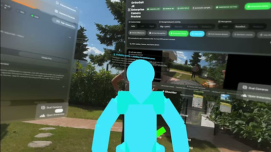
A. OrthoGait JR: Two-Camera 3D Fusion and Vision Pro Capture
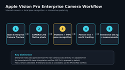
Two acquisition methods are shown in this video. The two-iOS workflow uses two calibrated external viewpoints and synchronized capture-time pairing for stereo triangulation. The Vision Pro Enterprise workflow uses approved internal headset-camera access for local pose recognition and immersive spatial presentation. If the white fused skeleton does not appear, first check the orange and green source streams, then shared-marker calibration, clock synchronization, full-body overlap, and camera geometry. Full setup guide:

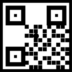
dowload the image and print in 12x12 cm both devices


TWO iOS CAMERAS, ONE FUSED 3D SKELETON
The stereo workflow uses two physical iPhone/iPad cameras viewing the same subject from different angles. Each device runs pose recognition and recognizes the same printed 12 cm shared QR calibration marker. OrthoGait JR uses the common marker transform, camera geometry, synchronized capture timestamps, and corresponding pose landmarks to reconstruct an accepted 3D pose.
Camera 1 appears in orange. Camera 2 appears in green. When synchronization, calibration, landmark confidence, stereo geometry, and ray-agreement gates pass, OrthoGait adds the white fused 3D skeleton.
Alt text: Diagram showing two iOS cameras viewing one subject, a shared 12 cm QR marker, orange and green source pose streams, and a white fused 3D skeleton in Apple Vision Pro.
SECTION 2 - PHYSICAL SETUP
For best results, place Apple Vision Pro and both iOS devices on the same trusted local Wi-Fi network and allow Local Network access.
In OrthoGait Capture, open Guided Capture and the pairing guide on each iPhone/iPad, select the same task, and start recognition.
Use the same printed 12 cm calibration marker for both cameras. Keep both cameras fixed after calibration.oan other way is in more menu to select advanced tool and prees. live mesurmentn by placing the shared qr code in same direction the shared should be recognized and this is proven by appearing in ar or in immersive space a green frame then automitcaly the camera is connected and by tap over star butoon in aplle vision is connected and streams to one window ui in case is not try to change the camera selection which by default is the first camera t
Maintain complete body visibility in both views. A practical starting point in an ordinary room is approximately 0.5-1.5 m separation between the two cameras with clearly different viewpoints and overlapping fields of view.
QUICK STEPS
1. Start Camera 1 and show the shared marker. Wait for the orange Primary source.
2. Start Camera 2 and show the same marker. Wait for the green Secondary source.
3. Keep the full subject visible in both views.
4. Wait for clock synchronization and capture-time pairing.
5. Confirm the white fused 3D skeleton appears after all stereo gates pass.
SECTION 3 - WHY CAMERA SYNCHRONIZATION MATTERS
Two packets that reach Vision Pro at the same moment were not necessarily captured at the same moment. Wi-Fi transport, pose inference, and device scheduling can add different delays to each camera.
OrthoGait JR Build 200 aligns each camera to the Vision Pro host clock using repeated four-timestamp synchronization exchanges. The system translates the real camera capture timestamp into a shared host timebase and pairs stereo observations by corrected capture time rather than network arrival order.
Alt text: Timeline schematic explaining camera capture time, network arrival time, Vision Pro host-clock correction, and synchronized stereo pairing.
SECTION 4 - WHAT THE COLORS MEAN
Orange: Camera 1 / Primary source pose.
Green: Camera 2 / Secondary source pose.
White: accepted fused 3D stereo skeleton.
Yellow: two streams may be present while triangulation is still pending.
Red: rejection or error in relevant diagnostic states.
Gray: idle or no current evidence.
The orange and green source rigs can remain visible for diagnostics even when the white fused rig is active.
SECTION 5 - IF THE WHITE FUSION DOES NOT APPEAR
Check both source streams first. Then confirm both cameras recognized the same 12 cm marker, clock synchronization is ready, the full body is visible in both views, and the two cameras have useful geometric separation. Fusion is intentionally withheld when the temporal or geometric quality gates fail.
Alt text: Troubleshooting flow for missing white stereo fusion, including camera connection, marker calibration, clock synchronization, body visibility, camera geometry, and quality gates.
Collapsible text is great for longer section titles and descriptions. It gives people access to all the info they need, while keeping your layout clean. Link your text to anything, or set your text box to expand on click. Write your text here...
Connect two iOS cameras
Shared 12 cm QR marker and camera placement
Capture-time synchronization
Stereo triangulation:
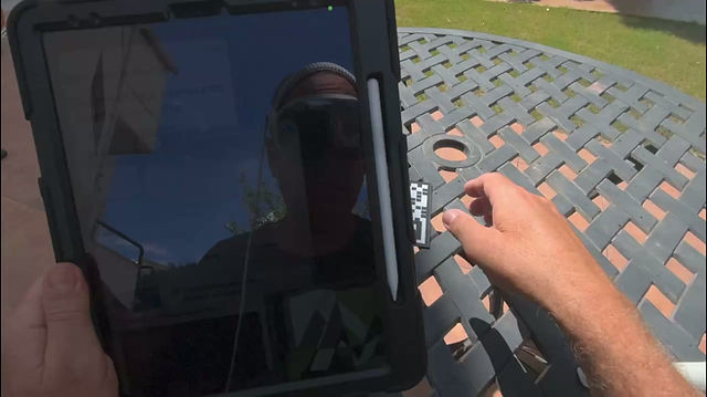
Two iOS cameras can be synchronized and calibrated to the same 12 cm QR marker so OrthoGait JR can triangulate accepted landmarks into one white 3D pose. The Enterprise build adds a separate Apple Vision Pro internal-camera workflow for local pose recognition and immersive review
 |  |  |  |
|---|---|---|---|
 |  |  |  |
 |
 |  |  |  |
|---|---|---|---|
 |
B. Enterpise Edition.
OrthoGait JR Entepize Version : Internal app vision Cameras open no other cameras need 3D Fusion and Vision Pro Enterprise Capture.