
FEMOROACETABULAR
IMPINGEMENT
APP
Femoroacetabular impingement (FAI) is a recognized cause of hip pain in the young adult and is postulated to be a major cause of early osteoarthritis. Femoroacetabular impingement is a mismatch between the shape of the head–neck junction and the acetabulum. Hump malformation of the femoral head-neck junction (Cam) or femoral head acetabular over-coverage (pincer) leads to a damage to the articular cartilage and arthritis. The best radiological means of measuring FAI is CT scan or MRI,

The app is medical software aimed for orthopaedic surgeons, providing tools that allow doctors to -Securely import medical images directly from the camera or stored photos. Offers a very convenient way to determine the most accurate possibly lines in order to measure the angles. By the aid of a circular transparent template, the points of interest are marked accurately. The circular template help to detect and mark easily, the alpha point where the radius of the curvature of the femoral head.

The app is a handy tool for an orthopaedic surgeon, radiologist, medical student or resident who wants objectively to monitor and determine femoroacetabular impingement (FAI) of the hip. The build-in comparison feature with the normal reference values and the combination of angles may help decide what could be considered normal or cam and or pincer type of FAI. The app is not a simple goniometer, is an enhanced product.This is particular useful especially in clinical settings where you need
Femoroacetabular impingement (FAI) is a recognized cause of hip pain in the young adult and is postulated to be a major cause of early osteoarthritis. Femoroacetabular impingement is a mismatch between the shape of the head–neck junction and the acetabulum. Hump malformation of the femoral head-neck junction (Cam) or femoral head acetabular over-coverage (pincer) leads to a damage to the articular cartilage and arthritis. The best radiological means of measuring FAI is CT scan or MRI,

Femoroacetabular impingement (FAI) is a recognized cause of hip pain in the young adult and is postulated to be a major cause of early osteoarthritis. Femoroacetabular impingement is a mismatch between the shape of the head–neck junction and the acetabulum. Hump malformation of the femoral head-neck junction (Cam) or femoral head acetabular over-coverage (pincer) leads to a damage to the articular cartilage and arthritis. The best radiological means of measuring FAI is CT scan or MRI, however, due to substantive radiation dose in CT and high cost in MRI, there are not routinely used in clinical practice for examinations. Femoral head/neck asphericity is best detected with the Dunn-projection-view in 45°, projection that is not always available. FAI is detectable also to an acceptable and conclusive level on standardised common AP pelvic radiographs.
The app is medical software aimed for orthopaedic surgeons, providing tools that allow doctors to:
-Securely import medical images directly from the camera or stored photos.
Offers a very convenient way to determine the most accurate possibly lines in order to measure the angles. By the aid of a circular transparent template, the points of interest are marked accurately. The circular template help to detect and mark easily, the alpha point - where the radius of the curvature of the femoral head first exits the circle of best fit corresponding to a circular head - real turning point of asphericity of femoral head
The drawn lines between points, allows app to estimate in radiographs, Center-Edge Angle (CE), α - angle (aA) and the anterior femoral offset ratio (AfOR). The measured values are compared with values from normal reference database. In case the measured angles are beyond the normal range, the hip is categorized as normal, dysplastic, borderline dysplastic hip and the type of femoroacetabular impingement (FAI) deformity namely cam type, pincer type or mixed is printed over the screen accordingly. Measures by the app are not affected by the X-ray projection.
-Save the planned images, for later review or consultation.
All information received from the software output must be clinically reviewed regarding its plausibility before patient treatment! The App is indicated for assisting healthcare professionals. Clinical judgment and experience are required to properly use the software. The software is not for primary image interpretation.
The app is a handy tool for an orthopaedic surgeon, radiologist, medical student or resident who wants objectively to monitor and determine femoroacetabular impingement (FAI) of the hip. The build-in comparison feature with the normal reference values and the combination of angles may help decide what could be considered normal or cam and or pincer type of FAI. The app is not a simple goniometer, is an enhanced product.This is particular useful especially in clinical settings where you need a quick results without losing time in looking for reference data and in doubtful cases.
Reference
1. Meyer DC, Beck M, Ellis T, Ganz R, Leunig M Comparison of six radiographic projections to assess femoral head/neck asphericity. Clin Orthop Relat Res. 2006 Apr;445:181-5.
2. Laborie LB, Lehmann TG, Engesæter IØ, Sera F, Engesæter LB, Rosendahl K.The alpha angle in cam-type femoroacetabular impingement: new reference intervals based on 2038 healthy young adults. Bone Joint J. 2014 Apr;96-B(4):449-54.
3. Ganz R, Parvizi J, Beck M, Leunig M, Nötzli H, Siebenrock KA (2003) Femoroacetabular impingement: a cause for osteoarthritis of the hip. Clin Orthop Relat Res 417:112–120
4. Mercier M, Dangin A, Ollier E, Bonin N. Does acetabular dysplasia affect outcome in arthroscopic treatment of cam femoroacetabular impingement? Case-control study with and without acetabular dysplasia.Orthop Traumatol Surg Res. 2019 Jan 7.. [Epub ahead of print]
5. Chegini S, Beck M, Ferguson SJ. The effects of impingement and dysplasia on stress distributions in the hip joint during sitting and walking: a finite element analysis. J Orthop Res. 2009 Feb;27(2):195-201.
6. Rubin DA (2013) Femoroacetabular impingement: fact, fiction, or fantasy? AJR Am J Roentgenol 2011(03):526–534
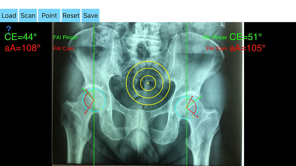
How to evaluate femoroacetabular impingement (FAI) with the App.
First you import medical images directly from the camera or stored photo. Aiming with the transparent circular template and by clicking the 'point' button the center of the femoral head is marked over the screen (C1 Point). Next by trying to fit to a best-fit circle to the contour of femoral head circumference. The radius of the circle is changed dynamically. Moving the attached finger you try to find the best fit circle to the contour of the femoral head. By pressing the 'point' button the C2 Point is marked and a yellow circle is drawn over the circumference of the femoral head.This will help to identify the alpha point. Next you aim to locate the left lateral acetabular edge and by clicking the 'point' button you mark the lateral acetabular edge (point E3). By moving the circular template you aim at the alpha point by pressing the point ‘Button’ the point A4 is marked. Alpha point is identified where the radius of the curvature of the femoral head first exits the yellow circle of best fit corresponding to a circular head-real, turning point of asphericity of femoral head. Next by aiming to locate the center of the femoral neck axis and pressing the ‘point’ button the N5 point is marked.
All information received from the software output must be clinically reviewed regarding its plausibility before patient treatment! The App is indicated for assisting healthcare professionals. Clinical judgment and experience are required to properly use the software. The software is not for primary image interpretation.
More help at www.orthopractis.com
In quick view, the following reference points you have to choose sequentially manually are shown below by the following order:
C1 → center of a femoral head.
C2 → dynamic cycle radius of femoral head.
E3 → lateral acetabular margin of acetabulum.
A4 → alpha point namely where radius of the curvature of the femoral head first exits the circle of best fit to femoral head.
N5 →center of femur neck canal axis.