
HandTremorLab
Observe hand motion. Review the signal. Understand its limits.


HandTremor — Tremor Lab
HandTremor is a hand-motion observation and clinical-support workspace for iPhone, iPad and Apple Vision Pro. It combines guided tasks, live hand landmarks, descriptive motion measurements and local session records. Its clinical guide and measurement reference explain what is measured, how it is calculated and why interpretation requires care.
HandTremor does not diagnose Parkinson's disease, essential tremor, dystonia or another medical condition. It does not classify diseases, estimate disease probability or recommend treatment. Clinical diagnostic accuracy has not been established. Consult a qualified healthcare professional for symptoms and medical decisions; do not change medication or delay care because of an app result.
CHOOSE THE EDITION FOR YOUR WORKFLOW
iPhone and iPad
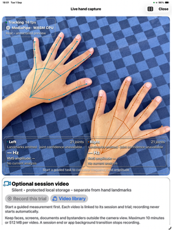
Use the device camera to estimate visible hand landmarks with Apple Vision or the bundled MediaPipe hand-landmark engine. The camera workspace displays the selected engine, the hand overlay, tracking status, live descriptive values and recording status. Camera-derived amplitude is relative to the image and hand scale, not calibrated millimetres or depth. The iOS edition supports iOS/iPadOS 17 or later on compatible hardware; actual tracking and processing performance depend on the device and conditions.
Apple Vision Pro Standard
Enter the immersive measurement space to use native ARKit hand tracking. Arrange the measurement and task-guide windows beside the workspace, and review source-labelled measurements and quality warnings. The Standard edition supports visionOS 2 or later on compatible Apple Vision Pro hardware. It does not access the headset's restricted main-camera video feed.
An optional, separately paired iPhone can supply extracted camera landmarks and timing. Choose the camera engine on the iPhone and verify the intended device during pairing. The connection sends landmarks, not camera images or video. Native Vision Pro hand tracking does not require an iPhone, and pairing is not automatic.
Apple Vision Pro Enterprise
The Enterprise edition supports approved business workflows on compatible Apple Vision Pro hardware running visionOS 26 or later. It retains native hand tracking and offers a separately selected main-camera path only when Apple's applicable enterprise API approval, entitlement, valid license, provisioning and user permission allow it. It does not unlock every Enterprise capability.
Restricted main-camera functionality is for eligible private business distribution, not an ordinary public or unlisted App Store edition. An app label or a copied license cannot grant access. Organization policy, hardware and permission may keep the camera unavailable. Contact support about an approved organizational deployment.
GUIDED HAND-MOTION OBSERVATION
Ten guided tasks cover supported rest, activated rest, posture, wing posture, rest-to-posture transition, kinetic movement, spiral, functional pouring, loading and pinch. Visual instructions, countdown controls and optional spoken guidance support a consistent workflow. Use only comfortable, safe tasks and stop for pain or fatigue. Do not use hot liquid, unsafe loads or hazardous objects.
Live and saved results can include dominant frequency, amplitude, effective sample rate, interruptions and other descriptive signal measures when the data supports them. Short, ambiguous or interrupted tracks may have unavailable results. A quality indicator describes the recording, not disease severity. Intended movement can contribute to the measured signal.
Frequency ranges overlap between clinical conditions. A measured frequency or amplitude is not a diagnostic cutoff. A normal-looking display does not rule out disease, and an unusual-looking display does not establish one.
ONE LIVE TABLE AND OPTIONAL USER LIMITS
Open Live table from the workspace or All measurements · live table from Settings. All 18 measurements for both hands appear in one spreadsheet-style view with separate values, units, status/bounds and last-updated fields. Scroll horizontally and vertically. Vision Pro opens a separate resizable window; iPhone/iPad uses a large sheet. Context identifies source, engine, task, quality and input age. Stale, insufficient, no-range and unavailable states remain explicit. Previous values are not new observations, and missing data is not zero motion. Existing measurement cards and one-moment CSV/JSON snapshots remain available.
Green means a fresh, finite, comparable value is inside a meaningful technical or matching user-entered bound. Red means outside a disclosed bound; technical warnings override a user-reference pass. Text and icons accompany colors. Built-in technical bounds are at least 20 Hz effective sample rate and at most 15% dropout. These are engineering limits, not population-normal thresholds. No clinical normal ranges are supplied. Green does not establish health or accuracy; red does not establish disease.
Use Configure limits to enter a lower bound, upper bound or both for the exact metric, source, engine, task and unit shown. These local preferences persist across launches and are not participant-specific; both hands can use a matching context. Built-in technical checks remain independently active even when limits are configured for the same field. Limits do not change the analyzer, calibrate the device or become disease cutoffs. Review, remove or reset them in Configured measurement limits; removal/reset is confirmed and is separate from deleting sessions or videos.
ON-DEMAND TIMELINES AND INDIVIDUAL CHARTS
During an active trial, choose Start timeline and confirm. One shared collector requests a checkpoint per second for all 18 measurements/both hands, capped at 600 snapshots or 600 seconds in memory. Actual elapsed/wall capture times, scheduled seconds, missed ticks, gaps and new/reused analysis flags remain recorded. Delays do not create backfilled or invented catch-up snapshots. Reused analyses retain freshness labels and are not new observations. The table shows the latest 30 checkpoints, while Export includes the full retained run. Display refresh is separate from retention; neither is the hand-capture rate or a raw tremor waveform.
Open a chosen hand/metric chart individually. Vision Pro provides separate resizable spatial windows beside the mixed immersive workspace; iPhone/iPad provides chart detail views. Opening a window does not start capture or retention. Missing, stale and incompatible source/engine/task/unit/bounds are not joined into a false continuous trend. Insufficient motion estimates are omitted; sample-rate/dropout diagnostics may remain explicitly labelled in their own quality segments. Rolling windows may overlap, so points are not independent observations. The chart does not establish diagnosis, disease progression, treatment response or clinically meaningful change.
Stop timeline preserves its history in memory without stopping the measurement. Capture/trial stop also stops retention, and the duration/count bound stops rather than overwrites older entries. There is no automatic next-trial or relaunch activation. Review/export before confirming Clear timeline or Start new timeline; the process ending also removes the in-memory history. Session records, raw landmarks and videos are separate.
Export freezes the whole retained timeline before TXT/JSON format choice. Review and acknowledge the health-data notice before Save/Share or copying selectable preview text. Both formats retain full numeric precision, all 36 rows per checkpoint, dates/timing, new/reused flags, gaps/freshness/status, source/engine/task/units and reference/technical bounds. Participant/examiner identifiers and free text, notes, session/trial IDs and raw uptime are omitted; these omissions do not make dates and motion anonymous. Format changes and later capture do not refresh the frozen copy. Use an authorized destination; external copies remain outside the app's deletion controls.
Explicit sharing prepares a protected, backup-excluded temporary .txt or .json source with the matching file type for a system copy. Both formats share a private cache capped at 32 MiB per report and 64 MiB / 64 files. Closing/cancelling the report, clearing the timeline or deleting sessions/videos does not immediately erase a transfer source. Recognized files from an earlier app process become eligible for cleanup after 24 hours at a later initialization or export; current-process files remain and system purging may occur. This does not promise exact 24-hour deletion or erase clipboard/recipient copies. Save to Files does not populate this cache and remains an alternative when it is full. Review the app-specific privacy notice.
TWO LOCAL CAMERA-RECOGNITION OPTIONS
Apple Vision and MediaPipe are working, selectable hand-landmark engines. The bundled MediaPipe option uses Tasks Vision with an on-device WebAssembly CPU runtime and a bundled hand-landmark model. It is not Google's native iOS SDK. The app does not download a model at runtime or send camera frames to a cloud inference service.
These engines perform machine-learning recognition of visible hands; HandTremor has no generative-AI assistant, chatbot or generated clinical interpretation. MediaPipe does not provide per-joint clinical accuracy probabilities. Its emitted-landmark validity must not be read as “100% accurate.”
Native ARKit world-space joints and camera-derived 2D landmarks are different measurement sources. Running a camera engine on Vision Pro does not turn its output into calibrated 3D hand tracking. Keep source, engine, units, task setup and tracking conditions consistent when reviewing repeated observations; the sources are not clinically interchangeable.
OPTIONAL SILENT VIDEO AND SEPARATE REANALYSIS
Video recording is off by default. During an active iPhone/iPad camera trial or an authorized Enterprise camera trial, choose Record this trial and confirm that you have permission to record everyone visible. A visible indicator shows when recording is active. The app captures no microphone audio.
Videos are retained in protected local app storage, excluded from backup, until you delete them. Recording is limited to 10 minutes or 512 MB per clip and stops when the trial ends or the app backgrounds. Keep faces, bystanders, screens and confidential documents outside the frame. File protection is not a substitute for appropriate device security and organizational access controls.
The video library supports playback and explicit import of an authorized video file. Import creates a silent local copy, leaving the external original unchanged. Imported clips are limited to 10 minutes, 4096 pixels per side and 512 MB after import. The acquisition conditions and calibration of an imported file are not verified by the app.
Select a recorded task and a range of up to 60 seconds, choose Apple Vision or MediaPipe, and agree to pause live capture before reprocessing. Reanalysis creates a new, engine-labelled offline report. It does not overwrite the original video or live measurement results. Processing may be slower than playback, and missing or insufficient data remains identified as such. Resume live capture explicitly afterward.
Native skeleton tracking and the iPhone landmark-only companion connection cannot create camera video on Vision Pro. To review iPhone footage in the Standard edition, explicitly record it on the iPhone and transfer an authorized file yourself. There is no automatic iPhone video relay.
LOCAL RECORDS AND USER-CONTROLLED EXPORTS
Save coded session context, task results and retained landmark samples locally. Review records in Sessions and deliberately export CSV, JSON, raw landmarks or a clinical-support report. Video reanalysis reports have their own separate JSON export. These files may contain sensitive health-related observations: use coded identifiers and an authorized destination.
Tap the live window's toolbar export control to freeze the displayed values, statuses, source/engine/task/units, relevant timestamps/ages and applied reference bounds while capture continues. A scrollable sheet shows the frozen snapshot time, a CSV or JSON format picker and the health-data notice. Changing format keeps the same snapshot. Choose Continue to Files to close the sheet and select a destination, or Cancel to keep it unshared. Later live updates do not change the prepared snapshot. This action adds no automatic continuous frame log, includes no participant notes or raw device uptime, and does not automatically upload a file. A snapshot can still contain sensitive observations; review its status and destination before sharing.
There is no HealthKit integration, automatic cloud synchronization or centralized patient database in this release. Nearby companion sharing is separately enabled and encrypted. Support links and email open only when selected; a session is not attached automatically.
External providers, mail services and export destinations have their own privacy and retention practices. Deleting a local record cannot delete a previously exported copy or an external original. The app provides explicit local deletion for videos and linked reports, individual sessions and all local sessions/videos.
User reference limits are separate app preferences, not protected session archives. Deleting sessions/videos leaves these limits in place; use their separate confirmed removal/reset controls. Do not enter identifying information into numeric reference settings or assume an exported combination of measurements, context and timing is anonymous.
CONSENT, CLINICAL CONTEXT AND RESPONSIBLE USE
A launch notice is shown each time a new app process starts. Capture requires acceptance and the applicable system permission. Video recording requires an additional confirmation. Declining keeps capture off. Withdrawing launch consent stops capture, companion activity, guidance and reanalysis; saved records remain until you explicitly delete them.
App consent is not a substitute for patient consent, research ethics approval, employee recording authorization or a lawful basis for processing personal data. Clinical and human-subject research use requires the appropriate professional, institutional, privacy and regulatory governance.
The version 3.5 method/study reference retains 47 entries, maps all 18 live fields, and provides per-measurement study/range limitations, 19 numbered references and six educational clinical contexts. These contexts are not generated from participant values or presented as diagnoses. The operating guide explains the table, one-second history and TXT/JSON export alongside retained charts and snapshots. Literature and proposed study use do not establish this app's clinical validity. Synthetic demonstration data is explicitly labelled and is not a real recording or clinical evidence.
BEFORE A RECORDING
Support the device, use even lighting and keep the wrist and fingertips within view. Check the selected source, active engine, anatomical hand labels and units. Occlusion, camera movement, orientation, hand visibility and processing rate can affect the result. Stop and investigate a misaligned overlay, frozen landmarks or unexpected status before retaining a measurement.
HELP AND CONTACT
Website: https://www.orthopractis.com
HandTremor support: https://www.orthopractis.com/handtremorapp
Privacy Policy: https://www.orthopractis.com/privacy
Terms of Use: https://www.orthopractis.com/terms-of-use
Support email: info@orthopractis.com
For help, provide the app version, device and operating system, selected source/engine and a description of the problem. Remove identifying information from screenshots, reports or logs before sending them. Do not send patient records or recordings unless an appropriate secure, authorized support arrangement has been agreed.
Feature description prepared for HandTremor 3.5.0, build 350. This is unpublished replacement copy, not a live website change or approval guarantee. Availability depends on the edition, compatible hardware, permissions and approved distribution route.


 |  |  |  |
|---|---|---|---|
 |  |  |
Tremor Lab / HandTremor — Operating Guide
Descriptive hand-motion estimation, not diagnosis
1. Before you start
Use a supported physical device and review the launch notice. Check the privacy notice and terms, acknowledge them to enter, or decline to keep capture closed. The choice resets on every new app process. Settings → Withdraw this launch's consent stops capture, sharing, speech and reanalysis without deleting saved records.
No edition provides generative AI advice or disease classification. Local Apple Vision and MediaPipe models locate hands; they do not determine the cause of movement. Check with a qualified clinician before making medical decisions. Do not use this app as an emergency alarm or delay care for acute symptoms.
2. Choose the capture source
iPhone/iPad: open iOS hand capture. The large camera sheet includes an Expand control for full-screen capture. Select an available front/rear camera and check the actual active engine and on-screen status. A Simulator cannot supply a real camera measurement.
Vision Pro Standard: open the immersive measurement space for native ARKit hand tracking, or separately pair the intended iPhone for camera-derived landmarks. Both devices must use a compatible protocol-v7 release. Pairing and optional remembered trust are explicit; only landmarks and timing are sent, not live camera pixels or video.
Vision Pro Enterprise: native ARKit remains available. For the separate main-camera source, open Enterprise Main Camera, check license/capability status and grant the required permission. Explicitly start the camera. Apple approval, correct signing/license, organizational eligibility and compatible physical hardware remain required. A camera-derived 2D result is not a native 3D measurement.
3. Select the engine before a trial
Settings → Recognition engine offers Apple Vision and MediaPipe · WASM CPU. MediaPipe's runtime and model are bundled and run locally; no API key, cloud model or runtime download is used. Native ARKit hand tracking is independent of this camera picker. For the iPhone companion, select the camera engine on the phone.
Check Actually active and the capture header. Preparing, unavailable and no-active-engine states are not successful recognition. The selected engine is not silently substituted. End the current trial/video before changing engine. An engine switch starts a new capture stream; one trial must not mix processing paths.
4. Check framing, coordinates and laterality
Support the device, use even lighting and a plain background, and keep the wrist and all fingertips visible. Check that overlays follow the correct anatomical hand and lie on the actual fingertips/wrist at the center and edges of the preview. Front-camera mirroring is display-only; Left/Right describes anatomy, not screen position.
Camera landmarks are stored in unmirrored sensor coordinates with top-left origin, orientation and image dimensions. Preview cropping/rotation is applied separately. If the overlay is offset or the side is wrong, stop, check camera/orientation/visibility, retry or select the other engine. Do not record a visibly incorrect track. Physical camera alignment must be verified for the device and orientations used.
Stale iOS observations are cleared instead of being shown as live indefinitely. Low inference rate or a timeout may leave measurements unavailable even while video capture works. Repeated tracking loss is missing data, not zero motion.
5. Measure and read the live displays
Use coded participant/examiner identifiers. Choose a task, review positioning instructions and use visual/spoken cues as appropriate. Keep the setup consistent. The usual guided duration is 20–30 seconds; you can stop early for comfort or safety. The analyzer needs adequate duration and tracking continuity before values appear.
The camera view and measurement workspace show hand observations, source/engine, duration and rolling estimates. Review dominant frequency, amplitude, timing, dropout and technical quality. Advanced values are available in Settings. Preview waveforms can be display-normalized; do not read pixel height as physical amplitude.
Open Live table or All measurements · live table from the workspace or Settings. One spreadsheet-style view shows all 18 measurements for both hands, with separate values, units, status/bounds and last-updated fields. Scroll horizontally for the hand columns and vertically for the rows. Vision Pro opens a separate resizable table window; iPhone/iPad presents a large sheet. The existing All live measurements cards, individual charts and one-moment snapshot exports remain available. The table displays these fields when the analyzer can supply them:
| Group | Fields |
|---|---|
| Timing and acquisition | Analyzed duration, usable sample count, effective sample rate, sample-timing jitter |
| Frequency | Dominant frequency, frequency resolution, spectral width |
| Spectral description | Peak-neighbourhood power ratio, spectral regularity, oscillation constancy |
| Motion | RMS amplitude, robust peak-to-peak amplitude, RMS velocity, RMS acceleration, projected angular RMS |
| Technical quality | Mean tracking indicator, dropout rate, composite quality score |
Source, actual recognition engine, task, units, result timestamps/age and quality/status accompany the numbers. Live, stale, last-result, unavailable and insufficient states are distinct: a previous result is not a fresh sample, a blank is not zero, and an insufficient result is not reassuring. The normal rolling analysis timing and requirements still apply; opening another window does not make values available before enough usable observations exist. MediaPipe has no per-joint confidence probability, so that field remains explicitly unavailable rather than showing 100% accuracy.
Green in the live table means a fresh, finite, comparable value is inside a meaningful technical or user-entered bound. Red means it is outside a disclosed bound; a technical warning takes priority over a user-reference pass. Text labels and icons accompany the colors. Missing/no-range values are neutral; stale or non-comparable values are explicitly marked and do not become green. Default technical bounds are effective sample rate at least 20 Hz and dropout at most 15%; existing analyzer quality warnings and insufficient checks remain unchanged. These are engineering checks, not population-normal limits. Green does not establish health, and red does not establish disease.
You may configure lower and/or upper reference bounds for an individual metric and exact capture source, recognition engine, task and unit. No clinical reference ranges are populated by default. A camera-relative bound must not be applied to a millimetre metric, another engine or another task. User reference bounds do not calibrate the device, validate a measurement, change the analyzer, or become disease cutoffs. Review the stated units/context and the range's origin before relying on it.
Tap a status/bounds cell in the live table, or Configure limits beside a measurement card. The editor retains that metric's exact source/engine/task/unit context while live capture continues. Use Save or Cancel, or Remove these limits with confirmation. These local range settings persist across app launches. A matching value below its lower bound or above its upper bound can be highlighted as outside a user reference. A missing matching range does not mean within normal limits. Limits are not participant-specific; both hands can share the same matching context. Check the context after changing source/engine/task.
Displayed measurement values and comparison limits use a decimal point without thousands grouping. The bounds editor accepts the device's decimal separator or a point; do not enter thousands separators.
Configured measurement limits lists stored settings. Reset all configured limits requires confirmation and is separate from deleting sessions/videos, which does not clear these reference settings. Editing or resetting a range does not start/stop capture or change a measured value. Stale, paused and last-result values retain explicit status rather than becoming new live alerts.
Frequency is a spectral component in the configured band, not a disease identifier. Camera amplitude is relative to observed hand span; native world-space output uses different units and assumptions. Quality is a technical recording indicator, not symptom severity or diagnostic certainty. MediaPipe exposes no per-joint confidence probability, so emitted landmarks must not be read as 100% confidence.
In the live table or Timeline, charts and export, choose Start timeline during the current active trial and confirm. Opening a table or chart does not start capture or retention. The single shared collector requests one checkpoint per scheduled second, retaining all 18 fields for both hands, up to 600 snapshots or 600 seconds. It records actual elapsed and wall-clock capture times plus the scheduled second and missed-tick count. A delayed tick is not backfilled, interpolated or caught up with invented snapshots. If no new analysis has arrived, that checkpoint explicitly marks the reused analysis and its freshness; reused values are not new observations. Recorded history displays the latest 30 checkpoints, with every metric available by horizontal scrolling; Export contains the whole retained run. The 0.5-second live-display refresh is not the retention cadence or capture rate. Overlapping rolling-analysis estimates are not independent observations, raw camera frames or a 1 Hz tremor waveform.
Choose a hand and metric, then Open separate chart window on Vision Pro or Open live chart on iPhone/iPad. Each Vision Pro chart is a separate resizable window alongside the mixed immersive measurement workspace; iOS uses a detail view. Opening, closing or rearranging chart views does not start capture or a timeline. Charts do not join across missing/stale values, incompatible source/engine/task/unit context, changed reference bounds or other marked discontinuities. Insufficient motion estimates are omitted; fresh sample-rate/dropout values may still be plotted as explicitly insufficient-quality technical diagnostics, separated from other quality states. A gap is not zero motion, and a line or color is not a disease classifier.
Choose Stop timeline when enough data has been collected. Stopping it does not stop the measurement; stopping or cancelling capture/trial activity stops the timeline. The retained timeline remains available for review/export in the current process until explicitly cleared or the process ends. The next trial does not automatically resume it. Clear timeline and Start new timeline require confirmation before removing/replacing retained history; export first if you need to keep it. At the duration/count limit it stops instead of overwriting the oldest values. A new app process starts with no retained timeline. Existing session/raw-landmark archives remain separate.
Settings → Measurement reference and numbered sources explains every output, its calculation, units, interpretation, source-code location and limitations. The version 3.5 catalog retains 47 core-method entries and adds explicit mappings for the 18 live fields, per-measurement study-use and reference-range limitations, 19 numbered sources and six educational clinical contexts. These contexts are not selected from a participant's values and do not generate diagnoses. The technical appendix, evidence summaries and supplied reference resources describe proposed study use and remaining validation needs, not patient data or app-validation results. See UPGRADE_3_5.md for the current table/retention/export workflow; earlier upgrade notes remain historical.
6. Guided tasks and safety
Supported rest, activated rest, outstretched posture, proximal/wing-beating posture, rest-to-posture transition, kinetic movement, spiral/tracing, pouring, loading and precision pinch describe different acquisition tasks. They do not automatically implement a validated clinical scale or verify that the task was performed correctly.
Standardize posture, task pace, target distance, container/load and context before comparison. Kinetic movement includes intended motion. Pinch estimates a motion vector, not force. Use only safe loads/containers, avoid hot liquids and stop for discomfort, fatigue, dizziness, distress or unsafe movement. Maintain environmental awareness in the headset.
7. Optional silent video
During an eligible camera trial choose Record this trial and confirm the separate recording notice. Check the red recording indicator. Recording captures the camera scene without microphone audio and may include bystanders or confidential surroundings. Obtain permission and frame minimally.
Stop/save or discard with the visible controls. Ending the trial or entering the background stops recording; finalization may finish saving already captured frames. The app does not automatically restart an interrupted trial or video. Limits are 10 minutes or 512 MB; low space, format changes or encoding failures can stop it earlier. Verify the final saved status.
Native ARKit skeleton tracking and a landmark-only companion stream cannot produce a camera video on Vision Pro. Record iPhone footage on the iPhone. Enterprise can record its approved main-camera source when available.
8. Playback, import and reanalysis
Open Settings → Session videos and reanalysis, or a saved session's linked-video view. Import only a video you are authorized to use. Import creates a separate silent local copy and leaves the external file unchanged. Import limits are 10 minutes, 512 MB and 4096 pixels per side.
For reanalysis select a video, task, Apple Vision or MediaPipe, start offset and a 12-, 30- or 60-second range within the clip. Confirm Pause live capture & reprocess. Longer clips can be reviewed in separate ranges. The result is labeled offline and retains the original source/engine provenance. Processing speed is not acquisition frame rate; analysis uses media presentation times.
Original video and live measurements remain unchanged. Each reanalysis produces a separate report or a clear insufficient-data explanation. No hand detected is not a normal clinical result. The selected task and imported setup are operator-supplied, not verified by the software.
When orientation changes within a video, ordinary playback uses the initial display transform; reanalysis uses the stored per-frame orientation timeline. The file is compressed and some offered capture frames may have been dropped. Reanalysis cannot recover missing frames or native 3D joints from 2D footage.
9. Save, export and delete
Review results before saving a session. A working session is bounded to 10 completed trials and 60,000 raw samples. These are resource safeguards. Save or discard before beginning a new working session when a bound is reached.
Tap Export snapshot in the live-measurement toolbar to freeze that moment's displayed values and metadata while capture continues. The scrollable Export snapshot sheet shows Snapshot time under Frozen displayed snapshot, a File format picker with CSV and JSON, and the Export health-related measurements? notice. Changing the format does not take a newer snapshot. Review the notice and both hands' status, then choose Continue to Files; the sheet closes before file destination selection opens. Choose Cancel to keep the snapshot unshared. Later live screen updates must not change the prepared snapshot. The snapshot includes result/source/engine/task/unit context, freshness/status, relevant timestamps/ages and the reference bounds used for its flags. It does not create a continuous per-frame log and does not include participant notes or raw device-uptime values. A cancelled export does not stop capture or automatically share a file.
Tap Export in the live table or timeline toolbar to freeze the whole currently displayed retained run. Export timeline offers TXT and JSON, run time, retained duration/count and a bounded preview. Format changes do not take a newer snapshot. Review the health-data notice and acknowledge it before Save .txt/.json to Files, Share .txt/.json or copying selectable preview text. Save dismisses the report sheet before opening the destination picker; Close/Cancel makes no new external copy and cannot recall an already saved/shared copy. Both formats retain full numeric precision, all 36 rows per checkpoint, units, source/engine/task, freshness/quality/status, new-versus-reused analyses, tracking gaps and reference bounds. Timing includes actual wall/elapsed capture time, scheduled second, missed ticks and analysis-generation/input-age context. These are not sensor-acquisition timestamps or fabricated missing samples. Later capture and limit edits cannot rewrite the frozen report. JSON provides structured fields for downstream processing; it does not convert reused rows into independent samples. The existing one-moment CSV/JSON snapshot action is unchanged.
Timeline TXT and JSON omit participant/examiner identifiers and free text, notes, session/trial identifiers and raw device-uptime values. Dates, motion estimates and capture context can still be sensitive and potentially identifiable. Choose only an authorized Files destination or share recipient and protect their copies. The in-memory timeline is not a saved session and is lost when cleared or the app process ends; explicitly export before that point if retention is required. The preview displays only the first 24,000 characters for responsiveness; a saved/shared file contains the complete frozen report.
Share .txt/.json prepares a protected, backup-excluded app-private temporary source file with the actual matching file type for a system copy. Limits are 32 MiB per report and 64 MiB / 64 files for that shared cache. Closing/cancelling, clearing the timeline or deleting sessions/videos does not immediately erase a transfer source. Recognized files from an earlier app process become eligible for cleanup after 24 hours at a later launch or share preparation; current-process files are kept and system temporary-storage purging may occur. This does not promise deletion exactly at 24 hours or erase clipboard, system or recipient copies. Save to Files does not add a file to the share cache and remains available if it is full. See the privacy notice before disclosure.
Session export provides JSON, metrics CSV, raw-landmark CSV and Markdown. Video-library export provides separate derived JSON, not raw video. Timing exports use elapsed/clip-relative intervals rather than device uptime. Read source, engine, units, warnings and offline/synthetic labels. Protect exported copies; local deletion does not recall them.
Delete selected sessions or videos with confirmation, or use Settings → Delete all local sessions and videos. Video deletion removes its derived reports but does not erase independent live measurements. Withdrawal of launch consent is not deletion. App Offload can preserve data; Delete App removes the local container but not external copies.
10. Synthetic examples
The synthetic-signal control is an openly labeled way to inspect graphs, session workflows and exports without hardware. Every synthetic recording must remain identified as synthetic. It is not a real hand recording, clinical evidence or a hidden review-only mode. Use actual authorized device capture for accuracy and performance evaluation.
11. Troubleshooting
Permission denied: review the relevant app permission in system Settings, then retry. The app cannot bypass Apple's permission prompts or Enterprise licensing.
No hands or poor alignment: check framing, lighting, camera choice, hand side and actual engine. Stop and retry; compare engines in separate trials. Never treat frozen or misaligned landmarks as valid data.
Low rate or unavailable metrics: reduce unrelated device load, improve visibility and allow a stable continuous trial. There is no guaranteed frame rate for MediaPipe CPU processing. Do not lower quality safeguards to obtain a desired clinical answer.
Red values or a stale/last-result label: read the specific technical warning or matching user-reference range and its units. Improve capture for technical warnings; check source/engine/task/unit and the entered bounds for user-range warnings. Red is not a disease alert, and a previous or unavailable value cannot establish a current clinical state.
Empty or stopped chart: opening a window does not activate timeline retention. Start an actual trial and explicitly start the timeline; allow the analyzer to produce adequate data. Check whether it stopped because the trial ended, capture was interrupted or the 600-snapshot/10-minute bound was reached. Repeated old results and gaps are not fresh chart points. Export a retained timeline before clearing it to start another. No timeline is restored after the app process exits.
Enterprise restart: close/reopen the appropriate capture workspace and start a fresh run. If camera authorization is unavailable, use native tracking only as an explicitly selected separate source; its output must not be relabeled camera data.
Speech unavailable: turn spoken guidance off and use visual cues. Video records no microphone audio. Apple console warnings alone do not establish the cause; report what actually failed on screen.
12. Support
Send the app version, device/OS, edition, source/engine, visible error and reproduction steps to info@orthopractis.com (mailto:info@orthopractis.com). Remove identifying details from examples. Public support email is not an approved patient-record transfer channel.
Website (https://www.orthopractis.com) · HandTremor support (https://www.orthopractis.com/handtremorapp) · Privacy Policy (https://www.orthopractis.com/privacy) · Terms of Use (https://www.orthopractis.com/terms-of-use)


Clinical evidence and interpretation policy
1. What can honestly be concluded
HandTremor estimates observed hand motion under a recorded setup. It reports descriptive frequency, displacement, derivative, projected-angle, rhythm and acquisition-quality quantities. It does not diagnose or exclude Parkinson disease, essential tremor, dystonic tremor or another condition, output disease probabilities, assign a clinical scale, or recommend treatment.
This is an actual intended-use boundary, not an attempt to rename an unvalidated health measurement as a wellness feature. No clinical or device-specific health-accuracy claim has been established for this build. App Review acceptance or a particular regulatory classification is not guaranteed by a disclaimer, literature list or redesigned interface.
There is no generative AI workflow. The Apple/MediaPipe options estimate landmarks on-device; the metrics and explanatory narrative are deterministic calculations and rules. Existing computer vision still involves model-based estimation and is not error-free.
2. Evidence map: source finding versus this implementation
| Number | Evidence type and relevant finding | Permitted inference for this project | Not established |
|---|---|---|---|
| [1] | Original MDS consensus classifies tremor using clinical characteristics, context and etiology | Preserve activation condition and associated context; avoid a diagnosis from one feature | Validation of camera metrics or a disease cutoff |
| [2] | Original MDS Parkinson criteria require a clinical syndrome and assessment beyond isolated oscillation | Hand-motion estimates cannot implement a complete Parkinson assessment | Disease probability, screening sensitivity or diagnostic status |
| [3] | Original comparison of Apple Vision/MediaPipe with reference methods distinguishes frequency performance from amplitude limitations | Validate frequency and amplitude separately; document geometry and engine | Accuracy of this app, this device, its WASM pipeline or its relative units |
| [4] | Original smartphone-video study compares frequency with a reference transducer | Use independent synchronized reference measurements in evaluation | This app's amplitude, regularity or quality thresholds |
| [5] | Original TETRAS reliability study concerns a clinical rating instrument | Keep clinical rating separate from landmark-derived metrics | An automatic landmark-to-TETRAS mapping |
| [6]–[9] | Official Apple/Google interfaces describe observations, acquisition and output structures | Identify APIs and preserve their metadata/limitations | Clinical efficacy, error guarantees or approval |
| [10] | Original MediaPipe technical preprint describes its model pipeline | Explain what kind of computer vision is used | This CPU/WebKit integration's performance |
| [11] | Original signal-processing paper explains data-window tradeoffs | Explain leakage and finite-record spectral resolution | Validation of custom disease or quality scores |
The clinical consensus uses history, activation conditions, distribution and other findings rather than one frequency alone. [1] (https://pubmed.ncbi.nlm.nih.gov/29193359/) The Parkinson criteria include bradykinesia with rest tremor or rigidity, followed by other clinical criteria; this application measures neither a complete syndrome nor all those findings. [2] (https://pubmed.ncbi.nlm.nih.gov/26474316/)
Wolke and colleagues studied 20 patients plus simulated movement and reported more encouraging peak-frequency findings than amplitude agreement. The paper explicitly limits clinical use of the tested amplitude estimates. These are findings about their pipeline, reference and setup—not a transfer of accuracy to HandTremor. [3] (https://pmc.ncbi.nlm.nih.gov/articles/PMC12008214/) Williams and colleagues' video-frequency study provides additional motivation for external reference comparison, not clinical certification of this code. [4] (https://pubmed.ncbi.nlm.nih.gov/34853806/)
3. What the outputs mean
Frequency describes the strongest analyzed peak. It can be affected by voluntary movement, aliasing, recording length, tracking noise and gaps. The2–15 Hz default search band and its sampling-dependent upper limit are engineering settings, not disease boundaries. “No peak” does not mean “no disease.”
Camera amplitude is % hand span in this implementation; there is no measured depth or validated calibration workflow. Native ARKit geometry is converted from metres to millimetres, but that unit conversion alone does not establish physical accuracy. Results from different sources/engines or camera setups must not be treated as interchangeable.
Velocity and acceleration are numerical derivatives of tracked displacement, not separate sensor measurements. Angular RMS describes a projected XY orientation, not clinical joint range of motion. Oscillation constancy is relative to the recording's own maximum, not a percentage of time living with symptoms.
Spectral regularity combines peak concentration and normalized entropy. Quality combines duration, confidence/validity, continuity and peak concentration. Both are custom developer-defined summaries. Their percentages are not confidence in a diagnosis, percent accurate, severity or clinical utility.
MediaPipe does not supply a per-joint location probability through the integration's chosen result interface; emitted validity is not 100% confidence. The confidence contribution to quality is withheld. Native ARKit fidelity weights also are application heuristics, not Apple-provided error probabilities. See the technical appendix (TECHNICAL_APPENDIX.md) and the 47-entry measurement catalog (Resources/Documentation/measurement_catalog.json) for exact source-derived formulas.
4. Internal limits versus diagnostic cutoffs
| Current numerical limit | Purpose | Must not be claimed |
|---|---|---|
| Minimum 3 seconds, 20 usable points, 32 resampled points | Permit numerical analysis | Minimum observation needed to diagnose disease |
| Search2…min(15, 0.45fs) Hz | Limit analyzed spectrum | Disease-specific normal/pathological range |
| Below20 Hz achieved cadence, gaps> 0.5 seconds or dropout> 35% | Mark technically weak captures insufficient | Clinical sensitivity or specificity threshold |
| Quality boundaries0.48/0.68/0.85 | Select internal display labels | Probability that a result is correct |
| Regularity labels0.3/0.6 | Select descriptive words | Dystonia or functional-tremor detection |
| Rest/action ratio> 1.5 or< 0.67 | Select task-comparison wording | Parkinson or essential-tremor classification |
| Matched laterality difference≥ 25% | Show a descriptive difference | Pathological asymmetry or meaningful change |
| Constancy windows≥ 25% of own maximum | Summarize relative persistence | Validated clinical constancy scale |
| Spectral floor1e−9 scaled units | Suppress numerical residue | Physical or clinical detection limit |
Frequency-context wording also uses < 4, 4–< 8, 8–< 12 and≥ 12 Hz categories. These categories are educational UI wording, not diagnostic rules. The app does not evaluate disease criteria, train a disease classifier or claim that irregularity detects dystonia. A favorable quality label does not convert a descriptive metric into a validated clinical endpoint.
5. Matched comparisons and context
Rest/action comparisons group by hand, source, recognition engine and unit. Left/right comparisons group by task, source, engine and unit. This prevents some inappropriate pooling, but does not control exact camera pose, hand posture, fatigue, medication timing, distance, load or time of acquisition.
Record relevant setup/context and repeat conditions before drawing conclusions. Defaults of20 or 30 seconds are workflow choices; the final live summary ordinarily represents only the last 12 seconds. This is why saved full raw trials and video can be useful for review while still requiring a defined analysis plan.
Entered context is not verified. Task instructions do not prove adherence. The rest-to-posture cue does not calculate re-emergent latency. No bilateral coherence, EMG activation, grip force, rigidity, bradykinesia decrement, automatic UPDRS/TETRAS score or validated clinical-change threshold is implemented. TETRAS has its own clinical evidence; no scale items or automatic scoring are reproduced here. [5] (https://pubmed.ncbi.nlm.nih.gov/23032792/)
6. Validation work still required
A defensible evaluation should define the intended population, tasks, devices/OS versions, engines, units, failure criteria and reference system before collecting data. The following are separate questions:
1. Analytical agreement: compare time, frequency and amplitude against synchronized reference acquisition with known limits; report bias, error distribution, agreement limits and failure rates.
2. Reproducibility: repeat within/between days, camera positions, raters, hands, lighting and relevant device thermal conditions.
3. Source/engine comparability: test native ARKit, Apple Vision, MediaPipe, companion and replay independently; do not infer equivalence from a shared graph.
4. Clinical validity and utility: prespecify outcomes, appropriate clinical reference and population; evaluate errors and consequences. Engineering quality labels are not a substitute.
5. Data integrity: verify timestamps, source locking, side identity, orientation, consent/background behavior, recording drops, import provenance and export-relative timebases.
This is a proposed validation framework, not a completed study or a claim of performance. The referenced publications cannot substitute for evaluation of the delivered implementation. Keep the physical-device and clinical-validation boundary visible in release materials and support documentation.
7. Structured study context
Metric-level study suggestions are proposed study-design uses, not evidence that a publication validated this endpoint or this app. Clinical-context cards are educational differential considerations, not diagnoses generated from a participant’s values. One-second exported rows retain actual timing, repeated-analysis flags and explicit missingness. Green/red identifies a scoped comparison, not a medical classification. The app never assigns a disease from a frequency band or custom quality/regularity score.
Rest-predominant oscillation
Observed context: A clinician observes oscillation at supported rest, possibly asymmetric, and compares matched rest and action recordings.
Relevant measurements: Dominant frequency, RMS amplitude, peak-to-peak amplitude and matched task/laterality comparisons; retain task, side, units and acquisition quality.
Possible clinical considerations: Parkinsonian tremor may be considered in the differential; isolated rest tremor and other tremor syndromes can overlap. This pattern is not a Parkinson diagnosis.
Required assessment: Examine bradykinesia, rigidity, gait and other neurological findings; apply the complete clinical criteria, medication history, exclusion criteria and red flags. The MDS motor-parkinsonism definition requires bradykinesia plus rest tremor or rigidity [2].
Not measured: Rigidity, validated bradykinesia decrement, diagnostic criteria and disease probability are not measured by this app.
Sources: [1], [2], [17].
Postural or kinetic oscillation
Observed context: Oscillation is observed during an outstretched posture or movement task; compare repeated recordings under a matched setup.
Relevant measurements: Task-specific frequency, RMS/peak-to-peak amplitude, spectral width and constancy; record whether voluntary movement or camera motion contaminated the task.
Possible clinical considerations: Essential tremor, enhanced physiological tremor, medication-related tremor, dystonic tremor and other causes may enter the clinical differential. Frequency alone cannot choose among them.
Required assessment: Document onset, duration, distribution, functional impact, associated signs and exposures. The 2018 ET syndrome includes bilateral upper-limb action tremor for at least three years and absence of other neurological signs meeting another syndrome [1]; the app does not establish those conditions.
Not measured: Long-term syndrome duration, thyroid/metabolic state, medication causality and a clinical tremor scale are not inferred.
Sources: [1], [5], [17].
Task- or position-dependent movement
Observed context: A clinician observes abnormal patterned postures, a position-dependent or jerky movement, a null point or an alleviating maneuver.
Relevant measurements: Retain task-specific amplitude, projected angular RMS, frequency and source-matched video for review. Document the observed posture separately.
Possible clinical considerations: Dystonia with tremor or tremor associated with dystonia can be considered when appropriate clinical signs are present. A broad peak or a low regularity score is not evidence sufficient to identify dystonia.
Required assessment: A movement-disorder examination should establish the phenomenology, distribution and associated findings using current dystonia terminology [12].
Not measured: Muscle contraction, co-contraction, a validated dystonia scale, sensory-trick detection and full 3D joint rotation are not measured.
Sources: [1], [12].
Marked variability or task-related change
Observed context: Frequency or amplitude appears to vary substantially between well-controlled tasks or recordings.
Relevant measurements: Inspect timing, gaps, source/engine changes, quality, raw/video context and repeated trial summaries before treating variation as biological.
Possible clinical considerations: Functional tremor can be one clinical consideration when positive clinical signs support it; tracking failure, voluntary movement and other tremors also vary. Irregularity alone must not produce a functional diagnosis.
Required assessment: Clinical distractibility/entrainment testing and, where indicated, a validated electrophysiological battery require specialist assessment [13].
Not measured: The validated functional-tremor test battery, EMG coherence, entrainment and its diagnostic accuracy are not implemented.
Sources: [1], [13], [14].
Movement near a target
Observed context: A clinician observes an increase in oscillation near a target during directed movement, with or without other coordination signs.
Relevant measurements: Kinetic-task amplitude and frequency may describe observed motion; compare like-for-like movement speed and target task while reviewing video.
Possible clinical considerations: Cerebellar/intention tremor and other kinetic tremors may be considered clinically. A low dominant frequency or high acceleration alone cannot identify a cerebellar disorder or lesion.
Required assessment: Examine dysmetria, gait, eye movements and associated neurological signs; investigate causes clinically. Sudden new tremor with weakness, speech change or severe imbalance warrants urgent medical assessment.
Not measured: Target-relative dysmetria, ataxia scale, lesion localization and diagnostic imaging findings are not calculated.
Sources: [1], [17].
Rest-to-posture time course
Observed context: Review an observed change from supported rest to sustained posture and any delay before oscillation is visible.
Relevant measurements: Use source timestamps, the recorded raise-arms cue and authorized raw/video review. A trend of 12-second overlapping estimates is not a direct latency trace.
Possible clinical considerations: Re-emergent tremor can occur in Parkinson disease, but the presence, timing and clinical context require independent assessment; a delayed plotted estimate may instead reflect analysis-window or processing delay.
Required assessment: Measure cue, actual posture transition and tremor onset with a separately validated protocol; interpret with the neurological examination [18].
Not measured: Validated onset detection and re-emergent latency are not implemented; the timeline must not be used as their substitute.
Sources: [1], [2], [18].
8. Evidence limitations and planned studies
The functional-tremor electrophysiological battery [13] and Tremor Stability Index [14] are different methods that this app does not implement. Their diagnostic-performance numbers must not be borrowed. The rest-to-posture cue is not validated latency measurement [18]. Apply verification, analytical and clinical validation separately [15], assess agreement rather than correlation alone [16], and prospectively report any future diagnostic study using appropriate reporting standards [19]. These are research-planning proposals, not completed validation.
References
[1] Bhatia KP et al. Consensus Statement on the classification of tremors. Movement Disorders. 2018;33:75–87. doi:10.1002/mds.27121. (https://pubmed.ncbi.nlm.nih.gov/29193359/) Original professional-society consensus. Supports describing activation condition and associated context rather than inferring a diagnosis from one motion feature. It does not validate this app.
[2] Postuma RB et al. MDS clinical diagnostic criteria for Parkinson's disease. Movement Disorders. 2015;30:1591–1601. doi:10.1002/mds.26424. (https://pubmed.ncbi.nlm.nih.gov/26474316/) Original professional-society diagnostic criteria. Explains why measured hand oscillation alone cannot establish Parkinson disease. The app does not implement these clinical criteria.
[3] Wolke R et al. Validity of tremor analysis using smartphone compatible computer vision frameworks. Scientific Reports. 2025;15:13391. doi:10.1038/s41598-025-97252-4. (https://pmc.ncbi.nlm.nih.gov/articles/PMC12008214/) Original experimental validation study. External Apple Vision/MediaPipe study found more encouraging frequency than amplitude agreement; its different acquisition and processing cannot be treated as validation of this build.
[4] Williams S et al. Accuracy of Smartphone Video for Contactless Measurement of Hand Tremor Frequency. Movement Disorders Clinical Practice. 2021;8:69–75. doi:10.1002/mdc3.13119. (https://pubmed.ncbi.nlm.nih.gov/34853806/) Original experimental validation study. Motivates comparison of video-derived frequency against a reference transducer; does not establish this app's accuracy or validate camera amplitude.
[5] Elble R et al. Reliability of a new scale for essential tremor. Movement Disorders. 2012;27:1567–1569. doi:10.1002/mds.25162. (https://pubmed.ncbi.nlm.nih.gov/23032792/) Original clinical rating-scale study. TETRAS is a separate clinical assessment instrument. This app neither reproduces its items nor automatically maps landmarks to a TETRAS score.
[6] Apple Developer Documentation: VNDetectHumanHandPoseRequest. (https://developer.apple.com/documentation/vision/vndetecthumanhandposerequest) Platform API documentation. Defines the camera hand-pose request and its landmark observations; API availability is not clinical measurement validation.
[7] Apple Developer Documentation: HandTrackingProvider. (https://developer.apple.com/documentation/arkit/handtrackingprovider) Platform API documentation. Defines native visionOS hand-anchor acquisition. Tracked world geometry is not a certified motion-capture accuracy claim.
[8] Apple Developer Documentation: CameraFrameProvider. (https://developer.apple.com/documentation/arkit/cameraframeprovider) Platform API documentation. Defines camera-image delivery and support/authorization boundaries used by the separately licensed Enterprise path.
[9] Google AI Edge: Hand landmarks detection guide for Web. (https://developers.google.com/edge/mediapipe/solutions/vision/hand_landmarker/web_js) Official SDK documentation. Documents video timestamps, handedness and landmarks. This build uses a bundled WebAssembly CPU runtime, not the native iOS SDK; no per-joint confidence probability is returned by its selected result interface.
[10] Zhang F et al. MediaPipe Hands: On-device Real-time Hand Tracking. 2020. arXiv:2006.10214. (https://arxiv.org/abs/2006.10214) Original technical preprint. Describes the palm-detector/landmark-model approach. It is neither a generative language model nor evidence of this app's clinical accuracy or CPU/WebKit throughput.
[11] Harris FJ. On the use of windows for harmonic analysis with the discrete Fourier transform. Proceedings of the IEEE. 1978;66:51–83. doi:10.1109/PROC.1978.10837. (https://doi.org/10.1109/PROC.1978.10837) Original signal-processing paper. Technical basis for explaining windowing/leakage and finite-record spectral resolution, not validation of the app's custom regularity or quality scores.
[12] Albanese A et al. Definition and Classification of Dystonia. Movement Disorders. 2025. doi:10.1002/mds.30220. (https://pmc.ncbi.nlm.nih.gov/articles/PMC12273609/) Original international consensus update. Provides current dystonia terminology and clinical phenotyping context. Patterned movement/postures require clinical examination; classification is not a camera-based diagnostic algorithm.
[13] Schwingenschuh P et al. Validation of laboratory-supported criteria for functional (psychogenic) tremor. Movement Disorders. 2016;31:555–562. doi:10.1002/mds.26525. (https://pubmed.ncbi.nlm.nih.gov/26879346/) Original prospective electrophysiological validation. Evaluates a specified multi-test electrophysiological battery. Its thresholds, sensitivity and specificity cannot be assigned to this app's camera regularity, confidence or timeline.
[14] di Biase L et al. Tremor stability index: a new tool for differential diagnosis in tremor syndromes. Brain. 2017;140:1977–1986. doi:10.1093/brain/awx104. (https://pmc.ncbi.nlm.nih.gov/articles/PMC5493195/) Original neurophysiological classifier study. The published Tremor Stability Index is distinct from HandTremor's spectral regularity, width and constancy. The app does not implement or claim the classifier's diagnostic performance.
[15] Goldsack JC et al. Verification, analytical validation, and clinical validation (V3): the foundation of determining fit-for-purpose for Biometric Monitoring Technologies. npj Digital Medicine. 2020;3:55. doi:10.1038/s41746-020-0260-4. (https://pmc.ncbi.nlm.nih.gov/articles/PMC7156507/) Original validation framework. Separates software/sensor verification, analytical agreement and clinical validity. A working export, a build pass or a green technical status satisfies none of these by itself.
[16] Bland JM, Altman DG. Statistical methods for assessing agreement between two methods of clinical measurement. Lancet. 1986;1:307–310. doi:10.1016/S0140-6736(86)90837-8. (https://pubmed.ncbi.nlm.nih.gov/2868172/) Original measurement-agreement methodology. Agreement must be assessed, not inferred from correlation. Define acceptable error before comparing engines/devices; account for repeated and overlapping observations in the study design.
[17] Haubenberger D et al. Transducer-based evaluation of tremor. Movement Disorders. 2016;31:1327–1336. doi:10.1002/mds.26671. (https://pmc.ncbi.nlm.nih.gov/articles/PMC5014626/) Professional-society task-force measurement assessment. Explains quantitative tremor measurement and its methodological limits. Instrument, task and analysis choices affect results; transducer evidence is not a validated normal range for optical hand landmarks.
[18] Jankovic J, Schwartz KS, Ondo W. Re-emergent tremor of Parkinson's disease. Journal of Neurology, Neurosurgery and Psychiatry. 1999;67:646–650. doi:10.1136/jnnp.67.5.646. (https://pubmed.ncbi.nlm.nih.gov/10519872/) Original clinical physiological study. Supports examining the rest-to-posture time course. The app records a cue but does not calculate validated re-emergent latency; one-second rolling summaries cannot reproduce that study's endpoint.
[19] Bossuyt PM et al. STARD 2015: an updated list of essential items for reporting diagnostic accuracy studies. BMJ. 2015;351:h5527. doi:10.1136/bmj.h5527. (https://www.bmj.com/content/351/bmj.h5527) Original reporting-guideline update. Any future diagnostic study must prespecify its index test, reference standard, population, thresholds and handling of failures, and report uncertainty. This release has no completed diagnostic-accuracy study.
