WhereMyCup App

The app offers a simplify study of the spinopelvic relationship for arthroplasty surgeons will effectively identify the complex “hip-spine” THA patient at high risk for postoperative instability

Evaluation of functional spinopelvic imaging in lateral radiographs in sitting and standing positions is of importance, especially in patient before total hip replacement surgery. Patients with concomitant hip and spine pathology undergoing primary total hip were identified as being at high risk for dislocation. Instability-prone patients arhtroplasty (THA) should appropriately assessed for the presence of deformity and abnormal spinopelvic mobility. The preoperative planning for THA should encompass evaluation of functional spinopelvic imaging in lateral radiographs in sitting and standing positions. Before planning cup position the surgeon should taken into account also anterior pelvic plane (APP) the coronal (functional) plane and the anterior pelvic plane tilt APPt or pelvic tilt and abnormal spinopelvic mechanics from standing to sitting.
 |  |  |
|---|---|---|
 |  |  |
 |  |  |
 |  |
The App is software aimed for orthopaedic surgeons, and allow to:
-securely import medical images directly from the camera or stored photos.
- mark certain points at the image of X-ray, and calculate at once the anterior pelvic plane tilt (APPt) or pelvic tilt , Pelvic incidence (PI), Sacral slope (SS), Lumbar lordosis (LL), Pelvic incidence Angle (PI) minus Lumbar lordosis Angle (LL)(PI–LL).
-to classify the patient into one of Four Categories of the Hip-Spine Classification (1A,1B,2A,2B) by integrating spinal alignment (spinal deformity defined at PI-LL mismatch >10 -Flatback deformity and mobility (stiffness- Defined as no change in Sacral Slope -SS between standing and sitting positions -ΔSS<10 deg )
-cases are categorised to group-specific recommendations for acetabular cup position respectively thus with minimal measurements will effectively identify the complex “hip-spine” THA patient at high risk for postoperative instability (1).
-save the planned images, for later review or consultation.The measured values are exported as txt file, ready to print or to input as cells to excel for research.
-in case of wrong selection powerful undo feature is present.
-According to measured parameters in standing and sitting lateral x- rays - based n a busy everyday practice, app offers a convenient way to perform radiographic measurements for spine, at the spinopelvic juncture - combing sitting and standing X-Rays, in a blink of an eye in front of your screen. The build in features of the app, allows results to be categorized and may help identify the complex “hip-spine” THA patient at high risk for postoperative instability and helps planning the optimum cup placement.
All information received from the software output must be clinically reviewed regarding its plausibility before patient treatment! App indicated for assisting healthcare professionals. Clinical judgment and experience are required to properly use the software.The software is not for primary image interpretation.
References
1. Luthringer, J.M. Vigdorchik A Preoperative Workup of a "Hip-Spine" Total Hip Arthroplasty Patient: A Simplified Approach to a Complex Problem. J Arthroplasty 2019 Jul;34(7S):S57-S70.
2. Pierrepont J, Hawdon G, Miles BP, et al. Variation in functional pelvic tilt in patients undergoing total hip arthroplasty. Bone Joint J 2017;99-B:184.
3 .Langston J, Pierrepont J, Gu Y, Shimmin A. Risk factors for increased sagittal pelvic motion causing unfavourable orientation of the acetabular component in patients undergoing total hip arthroplasty. Bone Joint J 2018;100-B:845.
4. Vigdorchik JM, Sharma AK, Dennis DA, Walter LR, Pierrepont JW, Shimmin AJ. The majority of total hip arthroplasty patients with a Stiff spine Do not have an Instrumented Fusion. J Arthroplasty 2020;35(6S):S252e4.
5. Esposito CI, Carroll KM, Sculco PK, Padgett DE, Jerabek SA, Mayman DJ. Total hip arthroplasty patients with Fixed spinopelvic Alignment are at higher risk of hip dislocation. J Arthroplasty 2018;33:1449.
6. Innmann MM, Merle C, Gotterbarm T, Ewerbeck V, Beaule PE, Grammatopoulos G. Can spinopelvic mobility be predicted in patients awaiting total hip arthroplasty? A prospective, diagnostic study of patients with end-stage hip osteoarthritis. Bone Joint J 2019;101-B:902.
7. DelSole EM, Vigdorchik JM, Schwarzkopf R, Errico TJ, Buckland AJ. Total hip arthroplasty in the spinal deformity Population: does degree of sagittal deformity affect rates of safe zone placement, Instability, or revision? J Arthroplasty 2017;32:1910.
8. Buckland AJ, Fernandez L, Shimmin AJ, Bare JV, McMahon SJ, Vigdorchik JM. Effects of sagittal spinal Alignment on Postural pelvic mobility in total hip arthroplasty Candidates. J Arthroplasty 2019;34:2663.
Introduction
 |  |
|---|---|
 |  |
 |  |
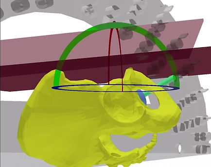
Anterior pelvic plane (APP) is defined by the plane between the two anterior superior iliac spines (ASISs) and the pubic symphysis on a lateral pelvic radiograph. On an imperfect lateral image where the ASISs are not superimposed, the superior end of the APP intersects the horizontal midpoint between the left and right ASIS.
The anterior pelvic plane tilt (APPt) is the measured angle created between the anterior pelvic plane (APP) and a vertical reference line. The anterior pelvic plane tilt APPt (or pelvic tilt) should be identified as being neutral (0), tilted posterior(-) or tilted anterior (+). In patients with no deformity, the APP will be vertical (neutral pelvic tilt) or parallel to the coronal (functional) plane of the body. Thus, the position of the cup in the functional standing position postoperatively will be the same as that referenced from the coronal plane of the body during cup implantation. As the implanted acetabular component and pelvis move as a single
unit, patients with posterior pelvic tilt will increase the functional anteversion of the cup while standing postoperatively. Conversely, anterior pelvic tilt will cause patients to functionally decrease cup anteversion in the standing position.
Sacral slope (SS) is defined at the angle subtended by a line parallel to the superior endplate of S1 and a horizontal reference line (HRL).
∆SS is the difference between the standing and sitting of SS-(Spinal stiffness Defined as no change in Sacral Slope -SS between standing and sitting positions -ΔSS<10)
Pelvic incidence (PI) is measured as the angle between a line drawn from the
center of the femoral heads to the center of the S1 endplate and a
second line drawn perpendicular to the S1 endplate
LL (also known as the lumbar lordotic angle) is the angle subtended by two lines drawn at the superior endplates of L1 and S1.
Pelvic incidence lumbar lordosis mismatch (PI-LL mismatch), PI-LL mismatch defined as PI minus LL) if is > 10 degree “flatback spinal deformity.”
These patients stand with posterior pelvic tilt, the APP is different from the FPP. It is crucial to recognize that this posterior pelvic tilt (of the APP) will increase the functional cup anteversion relative to the coronal plane while standing (FPP). After impaction, the cup and pelvis move as a single unit. If the cup is anteverted relative to the APP (bony landmarks), when the patient stands, it will tilt posteriorly with the pelvis, causing the face of the cup to open anteriorly, increasing anteversion relative to the coronal plane (FPP). Thus, anteversion targets must be referenced from the FPP (on the standing AP pelvis) and not from the APP. Failure to do so may result in excessive functional anteversion while standing and increase the risk anterior dislocation.
case example. Templated cup position of 40° of inclination and 20° of anteversion relative to the APP (or traditional bony landmarks intraoperatively) leads to functional cup position of 45° of inclination and 38° of anteversion when the patient stands. After accommodating for the patient's posterior pelvic tilt in the functional (standing) position, placement of the cup in 35° of inclination and 2° of anteversion relative to the APP will lead to a cup position of 40° of functional inclination and 20° of functional anteversion relative to the coronal plane when standing.

Anterior pelvic plane (APP) is defined by the plane between the two anterior superior iliac spines (ASISs) and the pubic symphysis on a lateral pelvic radiograph. On an imperfect lateral image where the ASISs are not superimposed, the superior end of the APP intersects the horizontal midpoint between the left and right ASIS. The anterior pelvic plane tilt (APPt) is the measured angle created between the anterior pelvic plane (APP) and a vertical reference line. The anterior pelvic plane tilt APP


Anterior pelvic plane (APP) is defined by the plane between the two anterior superior iliac spines (ASISs) and the pubic symphysis on a lateral pelvic radiograph. On an imperfect lateral image where the ASISs are not superimposed, the superior end of the APP intersects the horizontal midpoint between the left and right ASIS. The anterior pelvic plane tilt (APPt) is the measured angle created between the anterior pelvic plane (APP) and a vertical reference line. The anterior pelvic plane tilt APP
Patient is classified according to the Hip-Spine Classification for spinal deformity and spinal stiffnes
A:Normal spinal mobility (defined as >10 change in SS from
stand to sit).
B: Stiff spine (defined as <10 change in SS from stand to sit).
1.Normal spinal alignment (defined by PI-LL ± 10°).
2.Flatback deformity (PI-LL > 10° ).
Four Categories of the Hip-Spine Classification in THA and the necessary modification in Acetabular Position for each category.
1A. Normal alignment normal mobility normal, Normal anatomy and normal mobility (Anterior Pelvic Plane -APP similar to Functional Pelvic Plane-FPP) .
1B. Normal alignment stiff spine (<10° change in sacral slope from stand to sit). Stiff spine, needs more anteversion relative to the Anterior Pelvic Plane- APP which is similar to the Functional Pelvic Plane- FPP .Target 30° anteversion on the standing AP pelvis. Note the higher target due to the stiff spine to protect from a posterior dislocation.
2A. Flatback deformity (PI-LL>10°), normal mobility. Beware, the Functional Pelvic Plane-FPP is different from the Anterior Pelvic Plane-APP (posterior pelvic tilt from the spinal deformity causes more functional cup anteversion) Target 25-30 ° anteversion on the standing AP pelvis (to the functional pelvic plane)
2B. Flatback deformity (PI-LL>10°) stiff spine (<10° change in sacral slope from stand to sit) Functional Pelvic Plane- FPP is different from the APP and a stiff spine, so you need more anteversion, but be aware of the reference because the spinal deformity will cause more functional cup anteversion. Target 30° on the standing AP pelvis (to the Functional Pelvic Plane-FPP). Note the higher target due to the stiff spine to protect from a posterior dislocation. Most difficult to treat, very narrow window of safe zone. Consider dual mobility cup.
How to measure with the app:
At preoperative workup before using the app 2 radiographs should be taken: one standing lateral pelvis x-ray- including the lumbar spine and one sitting lateral pelvis x-ray- including the lumbar spine.
Once you load or capture the patient’s image by default the button “Std” is highlighted, meaning that you have to input preferably the standing X-ray to measure.
By clicking the button ‘point’ the over the two anterior superior iliac spines (ASISs) ( independent from site selection, left -right) and the pubic symphysis (point P1 ,P2 and P3 are marked) on a lateral pelvic radiograph respectively . the -anterior pelvic plane (APP) is drawn which is defined by the plane passing through the mid-distance of the two anterior superior iliac spines (ASISs) and the pubic symphysis (P3) on a lateral pelvic radiograph. The anterior pelvic plane tilt (APPt) is the measured angle created between the APP and a vertical reference line.
The anterior pelvic plane tilt APPt (or pelvic tilt) is being neutral (0), tilted posterior(-) or tilted anterior (+).
By clicking the button ‘point’ the point P4 over the left edge of the superior end plate of the L1 is marked and by the same manner locating the right edge of the superior end plate the point P5 is also marked- the order of marking does not affect the measurement. A green line P4,P5 over the anterior and posterior, superior endplate of vertebra body of L1 appears. With the transparent circular template you aim to locate the posterior corner on the top margin of S1 of the sacrum and by clicking the button ‘point’ the P6 point is marked and by clicking also the ‘point’, button at the anterior corner of the superior end plate of S1 vertebra , the point P7 is marked. Next you aim at center of the femoral head and by clicking the ‘point’ button you mark the center of femoral head (C8) .
Automatically the Pelvic incidence Angle (PI) ,Sacral slope Angle (SS) are anterior pelvic plane tilt (APPt) or Pelvic tilt Angle (PT), Lumbar lordosis Angle (LL),PI–LL Pelvic incidence Angle (PI) minus Lumbar lordosis Angle (LL) are being calculated and normal values appears respectively.
An asterisk appeared above of button Std meaning that the measured data are stored in ram and not discarded. By importing the new sitting X-ray the data from previous measurement namely of standing X-ray are not lost. Once you press the “Stt” button which is highlighted you can measure by selecting points in exact the same way.
With the transparent circular template you aim to locate the posterior corner on the top margin of S1 of the sacrum and by clicking the button “point” the P9 point is marked. Then by clicking the “point”, button at the anterior corner of the superior end plate of S1 vetebra, the point P10 is marked.
In case selecting wrong points by pressing the undo button the procedure backtrack one step each time before.
Finishing the measurements the asterisk appears above button “Stt” meaning that the measured data are stored in ram
The detailed analysis of data by comparing the standing and sitting position is exported and by pressing the save button the image and the relevant data are stored. The stored txt file can be open with notepad or numbers externally.
In quick view the points that should be selected:
In standing Xray Std * is highlighted
P1-> anterior superior iliac spine (Left or Right)
P2 ->anterior superior iliac spine ( Right or Left )
P3-> pubic symphysis
P4 -> posterior corner on the top margin of L1 vetebra.
P5-> anterior corner of the superior end plate of L1 vetebra
P6-> posterior corner on the top margin of S1 vetebra of the sacrum.
P7-> anterior corner of the superior end plate of S1 vetebra.
C8 :-center of the femoral head.
In sitting Xray Sit * is highlighted
P9 ->posterior corner on the top margin of S1 vetebra of the sacrum.
P10->anterior corner of the superior end plate of S1 vetebra.
Hip surgeons should consider these factors when planning for THA in this patient population, maximizing impingement-free range of motion with proper component position, large head size, offset, leg length, and possibly using a dual-mobility bearing.